


 Extract (6-MSITC) in Healthy Older Adults")
: An In-Depth Exploration into its Thermogenic Role and Social Significance")


The Problem
According to the CDC, 1.5 million women in the US are infertile.
The causes are many, but roughly a quarter of all cases are due to irregular or abnormal function of the ovaries.
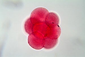
The ovaries are central organs for female reproduction because they release eggs that, once fertilized, develop into embryos.
The ovaries also act like conductors, releasing hormones that orchestrate the entire reproductive system to function as one dynamic unit to sustain fertility.
It’s a highly complex and fragile system that sometimes goes awry.
Age is a big factor. Although increasingly couples are waiting longer to have kids, it becomes much harder for women to get pregnant past the age of 35. Most scientists currently believe that women are born with all the eggs that they will ever produce (although recent research suggests otherwise).
With age, the quality of the eggs declines and may result in higher chances of acquiring random genetic mutations.
The ovary milieu — a niche of hormone-releasing cells and other structures that help nurture the eggs — also begins to decay, making it harder for ovaries to release eggs on schedule.
Childhood cancer is another big factor.
Chemotherapy drugs often damage a dividing cell’s DNA, which kills off cancer cells but also harms egg cells and increases the chance of infertility.
“One of the biggest concerns for patients diagnosed with cancer is how the treatment may affect their fertility and hormone health,” says Laronda.
We hope that the prosthetic implant helps to restore their quality of life, she says.
The Implant
In order for the implant to properly work, it needs three things: immature egg cells (called “oocytes”), hormone-releasing cells that support oocytes, and a flexible but rigid enough scaffold that can support the growth of both cell types.
To generate the scaffold, scientists turned to gelatin, a glutinous biomaterial derived from the skin of animals. (It’s also the main ingredient of Jell-O.)
Rather than recreating the entire complex structure of an ovary, the team took a minimalistic approach: using 3D printing, they generated scaffolds of different sizes and shapes to see which configuration works best.
Using immortalized human cells, Laronda and team found that the best structure had crisscrossing struts.
The design probably worked best because it has multiple anchor points for cells to latch onto and also has a sufficient amount of space for oocytes to grow, explained the team.
Finally, mouse ovarian follicles — ball shaped units that contain both the oocyte and supporting cells — were seeded onto the scaffold to complete the prosthesis, which was then implanted into female mice that had their ovaries surgically removed.
The first promising sign came when the mice restored their hormonal cycle, suggesting that at least the hormone-producing cells were alive and functional.
Just this one result would’ve been awesome. Many women have decreased production of reproductive hormones due to diseases such as PCOS (Polycystic Ovary Syndrome) that can cause problems with bone density, weight and cardiovascular health, explained the researchers.
A similar prosthesis may be able to combat those issues in at risk women.
But then came the bombshell: following the implant, the mice ovulated, gave birth to healthy pups and were able to nurse them until weaning.
When researchers examined the implant, they found that blood vessels had innervated the entire structure, thus providing nutrients and washing away waste and allowed the oocytes to develop normally into viable pups.
The fact that blood vessels automatically formed a network around the implant is huge. Often we need to add substances to stimulate the process, says Laronda, which could cause side effects and other complications.
This 3D printing manufacturing technique bypasses that requirement and could influence future work on complex soft tissue replacement, she says.
The Study
By removing a female mouse’s ovary and replacing it with a bioprosthetic ovary, the mouse was able to not only ovulate but also give birth to healthy pups.
The moms were even able to nurse their young.
The bioprosthetic ovaries are constructed of 3-D printed scaffolds that house immature eggs, and have been successful in boosting hormone production and restoring fertility in mice, which was the ultimate goal of the research.
“This research shows these bioprosthetic ovaries have long-term, durable function,” said Teresa K. Woodruff, a reproductive scientist and director of the Women’s Health Research Institute at Feinberg. “Using bioengineering, instead of transplanting from a cadaver, to create organ structures that function and restore the health of that tissue for that person, is the holy grail of bioengineering for regenerative medicine.”
The paper will be published May 16 in Nature Communications.
How is this research different from other 3-D printed structures?
What sets this research apart from other labs is the architecture of the scaffold and the material, or “ink,” the scientists are using, said Ramille Shah, assistant professor of materials science and engineering at McCormick and of surgery at Feinberg.
That material is gelatin, which is a biological hydrogel made from broken-down collagen that is safe to use in humans.
The scientists knew that whatever scaffold they created needed to be made of organic materials that were rigid enough to be handled during surgery and porous enough to naturally interact with the mouse’s body tissues.
“Most hydrogels are very weak, since they’re made up of mostly water, and will often collapse on themselves,” Shah said.
“But we found a gelatin temperature that allows it to be self-supporting, not collapse, and lead to building multiple layers. No one else has been able to print gelatin with such well-defined and self-supported geometry.”
That geometry directly links to whether or not the ovarian follicles, organized hormone-producing support cells surrounding an immature egg cell, will survive in the ovary, which was one of the bigger findings in the study.
“This is the first study that demonstrates that scaffold architecture makes a difference in follicle survival,” Shah said. “We wouldn’t be able to do that if we didn’t use a 3-D printer platform.”
How does this impact humans?
The scientists’ sole objective for developing the bioprosthetic ovaries was to help restore fertility and hormone production in women who have undergone adult cancer treatments or those who survived childhood cancer and now have increased risks of infertility and hormone-based developmental issues.
“What happens with some of our cancer patients is that their ovaries don’t function at a high enough level and they need to use hormone replacement therapies in order to trigger puberty,” said Monica Laronda, co-lead author of this research and a former post-doctoral fellow in the Woodruff lab.
“The purpose of this scaffold is to recapitulate how an ovary would function. We’re thinking big picture, meaning every stage of the girl’s life, so puberty through adulthood to a natural menopause.”
Laronda is now an assistant professor at the Stanley Manne Children’s Research Institute at the Ann & Robert H. Lurie Children’s Hospital.
Additionally, the successful creation of 3-D printed implants to replace complex soft tissue could significantly impact future work in soft tissue regenerative medicine.
Technically, how does biological 3-D printing work?
3-D printing an ovary structure is similar to a child using Lincoln Logs, said Alexandra Rutz, co-lead author of the study and a former biomedical engineering graduate fellow in Shah’s Tissue Engineering and Additive Manufacturing (TEAM) lab at the Simpson Querrey Institute.
Children can lay the logs at right angles to form structures. Depending on the distance between the logs, the structure changes to build a window or a door, etc.
“3-D printing is done by depositing filaments,” said Rutz, who is now a Whitaker International Postdoctoral Scholar at École Des Mines De Saint-Étienne in Gardanne, France.
“You can control the distance between those filaments, as well as the advancing angle between layers, and that would give us different pore sizes and different pore geometries.”
In Northwestern’s lab, the researchers call these 3-D printed structures “scaffolds,” and liken them to the scaffolding that temporarily surrounds a building while it undergoes repairs.
“Every organ has a skeleton,” said Woodruff, who also is the Thomas J. Watkins Memorial Professor of Obstetrics and Gynecology and a member of the Robert H. Lurie Comprehensive Cancer Center of Northwestern University. “We learned what that ovary skeleton looked like and used it as model for the bioprosthetic ovary implant.”
In a building, the scaffolding supports the materials needed to repair the building until it’s eventually removed. What’s left is a structure capable of holding itself up.
Similarly, the 3-D printed “scaffold” or “skeleton” is implanted into a female and its pores can be used to optimize how follicles, or immature eggs, get wedged within the scaffold.
The scaffold supports the survival of the mouse’s immature egg cells and the cells that produce hormones to boost production.
The open structure also allows room for the egg cells to mature and ovulate, as well as blood vessels to form within the implant enabling the hormones to circulate within the mouse bloodstream and trigger lactation after giving birth.
The all-female McCormick-Feinberg collaboration for this research was “very fruitful,” Shah said, adding that it was motivational to be part of an all-female team doing research towards finding solutions to female health issues.
“What really makes a collaboration work are the personalities and being able to find the humor in the research,” Shah said. “Teresa and I joked that we’re grandparents of these pups.”


