


 Extract (6-MSITC) in Healthy Older Adults")
: An In-Depth Exploration into its Thermogenic Role and Social Significance")


Scientists from Lancaster University in the UK have discovered that immune responses originally found to prevent fungal infections are also important in eliminating Trichinella spiralis, a round worm and the causative agent of Trichinosis.
People acquire trichinellosis by consuming raw or undercooked meat infected with the Trichinella parasite, particularly wild game meat or pork.
Depending on the classification used, there are several species of Trichinella: T. spiralis, T. pseudospiralis, T. nativa, T. murelli, T. nelsoni, T. britovi, T. papuae, and T. zimbabwensis, all but the last of which have been implicated in human disease.
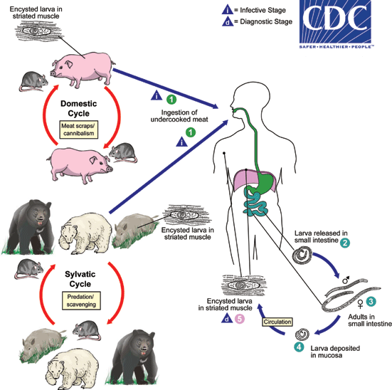
Adult worms and encysted larvae develop within a single vertebrate host, and an infected animal serves as a definitive host and potential intermediate host.
A second host is required to perpetuate the life cycle of Trichinella. The domestic cycle most often involved pigs and anthropophilic rodents, but other domestic animals such as horses can be involved.
In the sylvatic cycle, the range of infected animals is great, but animals most often associated as sources of human infection are bear, moose and wild boar.
Trichinellosis is caused by the ingestion of undercooked meat containing encysted larvae (except for T. pseudospiralis and T. papuae, which do not encyst) of Trichinella species (1).
After exposure to gastric acid and pepsin, the larvae are released from the cysts(2) and invade the small bowel mucosa where they develop into adult worms(3).
Females are 2.2 mm in length; males 1.2 mm.
The life span in the small bowel is about four weeks. After 1 week, the females release larvae(4) that migrate to striated muscles where they encyst(5) .
Diagnosis is usually made based on clinical symptoms, and is confirmed by serology or identification of encysted or non-encysted larvae in biopsy or autopsy specimens.
Geographic Distribution
Worldwide. Most common in parts of Europe and the United States.
Clinical Presentation
Light infections may be asymptomatic. Intestinal invasion can be accompanied by gastrointestinal symptoms (diarrhea, abdominal pain, vomiting).
Larval migration into muscle tissues (one week after infection) can cause periorbital and facial edema, conjunctivitis, fever, myalgias, splinter hemorrhages, rashes, and peripheral eosinophilia.
Occasional life-threatening manifestations include myocarditis, central nervous system involvement, and pneumonitis.
Larval encystment in the muscles causes myalgia and weakness, followed by subsidence of symptoms.
Consumption of contaminated meat contains “nurse cells” of the parasite.
Once in the stomach the “nurse cells” hatch releasing infective larvae which then bury themselves within the lining of the small intestine.
Previously immune responses to expel the parasite have been shown to rely on white blood cells called T-helper 2 cells, specialised for eliminating gastrointestinal parasites.
However, scientists at Lancaster discovered that following this T-helper 2 response, a second T-helper 17 response, previously shown to be specialised for eliminating fungal infections and certain bacterial infections occurred.
In collaboration with Professors Mark Travis and Richard Grencis from the University of Manchester, they were able to identify how these T-helper 17 cells arose and that they were key in maintaining the intestinal muscle contractions needed to flush out the worms.
The findings have been published in the journal PLOS Pathogens and show that mice lacking the ability to activate a key signalling molecule important in producing T-helper 17 cells have a reduced ability to expel the parasite.
Interestingly, they saw a delayed transit time in the small intestine hinting at alterations in muscle contraction.
In isolating the small intestine they demonstrated that a key molecule produced from T-helper 17 cells, termed IL-17, could increase intestinal contraction and restoring levels of this IL-17 in their mice rescued their ability to expel the parasite.
Dr. John Worthington from the Department of Biomedical and Life Science at Lancaster led the research:
“We were quite surprised by what we found during this study.
Normally, these immune responses are thought of as acting quite distinctly depending on what type of infection you may have.
It’s well established that the T-helper 2 response is beneficial during gastrointestinal worm infections, so traditionally any other response would be thought of as hindering worm expulsion.
So, it was quite surprising to see that this late acting T-helper 17 response was actually beneficial to the mouse’s ability to resolve an infection and get rid of the worm.”
Dr. Worthington continues: “Our study provides novel insights into how the immune system interacts with muscle contraction during intestinal inflammation.
Although the occurrence of this infection is very rare in the developed world, we hope it will help us to design new treatments for the many millions of people who suffer from intestinal parasitic infections worldwide and may even inform other intestinal diseases involving altered muscle function.”
More information:PLOS Pathogens (2019). DOI: 10.1371/journal.ppat.1007657
Journal information: PLoS Pathogens
Provided by Lancaster University


