


 Extract (6-MSITC) in Healthy Older Adults")
: An In-Depth Exploration into its Thermogenic Role and Social Significance")


The sense of touch is formed by a large number of tactile organs situated just under the surface of the skin between the epidermis and the dermis (figure 1).
The tactile organs are simple receptors connected by nerve axons.
The receptors sense pressure on the skin, and that is how you can feel touch.
There are tactile organs all over the body.
The distance between the receptors determines sensitivity, which differs for different parts of your body.
Fingers, tongue, lips, nose and forehead are very sensitive to touch, meaning that these parts have a higher density of tactile organs.
Not surprisingly, perhaps, these are all important parts of the body for feeling or touching.
Other areas, such as the back or the soles of the feet, have a lower density of tactile receptors and are less sensitive to touch.
It is necessary to use your other senses, such as movement or sense of temperature, if you want to know more than simply the fact that something touched you.
You can use your sense of movement to explore the object with your hands, to find out its shape, its external structure and so on.
Yet it is still difficult to determine what it might be. Your sight will give you the most information.
Your tactile sense delineates your body.
It indicates where you end and something else begins.
Without the sense of touch you would not feel this boundary, and you would not know where you stop being. You would literally be boundless and flow into the other.
Without a sense of touch, you would have no physical self-awareness.
A team of researchers at the Institute of Neurosciences of Alicante of the ISIC in Spain has found that the sense of touch arises in the brain before birth – at least in mice.
In their paper published in the journal Science, the group outlines their study of the embryonic stages of the development of the brain in mice and what they learned from it.
The development of a sense of touch has been studied by scientists for many years, but how it develops is still unclear.
Prior research has shown that once it has developed, it exists as a sort of map imprinted on the cerebral cortex.
Some have suggested that a basic map is created in the brain before birth and data points for it are added as newborns develop – sensory input from various body parts is simply added to the map.
But now, that view might have to change, as the team in Spain reports evidence that suggests the map is already in place by the time a baby is born.
In their efforts, the researchers used mice to better understand how the sense of touch might develop because mice have what are known as cortical barrels – regions within the cortical layer that are visibly darker when stained.
Prior research has shown that the map outlining the sense of touch in mice can actually be seen under a microscope by studying the cortical barrels.
The barrel cortex refers to a region of somatosensory cortex that is identifiable in some species of rodents and species of at least two other orders and contains the barrel field.
The ‘barrels’ of the barrel field are regions within cortical layer IV that are visibly darker when stained to reveal the presence of cytochrome c oxidase, and are separated from each other by lighter areas called septa.
These dark-staining regions are a major target for somatosensory inputs from the thalamus, and each barrel corresponds to a region of the body.
Due to this distinctive cellular structure, organisation, and functional significance, the barrel cortex is a useful tool to understand cortical processing and has played an important role in neuroscience.
The majority of what is known about corticothalamic processing comes from studying the barrel cortex and researchers have intensively studied the barrel cortex as a model of neocortical column.
The most distinctive aspect of the barrel field are the whisker barrels. These structures were first discovered by Woolsey and Van der Loos.
Staining in the whisker barrels is more distinct than that in other areas of somatosensory cortex.
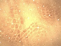
Pictomicrograph shows the barrel field in layer IV of the rat somatosensory cortex. Each barrel receives input from one whisker. The tissue in the image has been stained with cytochrome oxidase and is 50μm thick.
Recognizing that the array was similar to that of the vibrissae on the mystacial pad, they hypothesized that the barrels were the “cortical correlates of the mystacial vibrissae” and that “one barrel represents one vibrissa”.
Whereas small non-whisker areas of barrel cortex correspond to large and sometimes overlapping areas of the body, each much larger whisker barrel corresponds to a single whisker.
As a result, the whisker barrels are the focus of the majority of barrel cortex research, and ‘barrel cortex’ is often used to refer primarily to the whisker barrels. Consequently, much of this article focuses on rodent whisker barrel cortex.
Prior research has also shown that brain function such as recognizing and responding to sensory information arises due to electrical signals that stimulate growth of neurons.
Thus, to learn more about the development of the sense of touch in mice (at least concerning its whiskers) the researchers studied brain slices at various stages of development to monitor cortical barrel development, and also studied brain waves that have previously been identified as those associated with sensory processing.
In looking at their results, the researchers found that the sensory map created to process the sense of touch was built while the mice were still embryos.
And it developed due to signals sent to the cerebral cortex by the thalamus, which also plays a major role in relaying synaptic information throughout the life of the mouse.
The researchers suggest that it is likely the same process occurs in humans because “the organization of the cortex is conserved evolutionarily between species.”.
More information: Noelia Antón-Bolaños et al. Prenatal activity from thalamic neurons governs the emergence of functional cortical maps in mice, Science (2019). DOI: 10.1126/science.aav7617
Journal information: Science


