


 Extract (6-MSITC) in Healthy Older Adults")
: An In-Depth Exploration into its Thermogenic Role and Social Significance")


Although the exact causes of multiple sclerosis still remain unknown, it is assumed that the disease is triggered by a combination of genetic and environmental risk factors.
But which? In a mouse model of the disease, researchers at the University of Geneva (UNIGE) and the Geneva University Hospitals (HUG), Switzerland, studied the potential link between transient cerebral viral infections in early childhood and the development of this cerebral autoimmune disease later in life.
Indeed, the brain area affected by viral infection during childhood undergoes a change that can call, a long time later, on the immune system to turn against itself at this precise location, triggering autoimmune lesions.
These results, which are published in the journal Science Translational Medicine, provide a first step in answering one of the possible environmental causes of this serious disease..
Multiple sclerosis affects one in 1,000 people in Switzerland, two-thirds of whom are women.
It is the most common auto-immune disease affecting the brain.
Up to date, there is still neither a cure available nor a clear understanding of the factors that trigger this disease at around 30 years of age.
“We asked ourselves whether brain viral infections that could be contracted in early childhood were among the possible causes,” says Doron Merkler, a professor in the Department of Pathology and Immunology in UNIGE’s Faculty of Medicine and senior consultant in the Clinical Pathology Service of the HUG.
Such transient brain infections can be controlled quickly by the immune system, without the affected individual even noticing any symptoms.
“But these transient infections may, under certain circumstances, leave a local footprint, an inflammatory signature, in the brain,” continues the researcher.
The childhood: a pivotal moment influencing disease risk
The scientists induced a transient viral infection in a group of adult mice and in a group of mice at a very young age in order to test this hypothesis. Karin Steinbach, a researcher in the same department, explains:
“In both cases, the mice showed no signs of the disease and eliminated the infection within a week with a similar anti-viral immune response.”
The scientists then allowed the two groups of mice to grow older before they were transferred with self-reactive cells, which can target the normal brain structure and are also thought to contribute to the illness of patients with multiple sclerosis.
“These self-reactive cells are present in most of us, but do not necessarily induce a disease, since they are controlled by different regulatory mechanisms and usually don’t have access to the brain,” explains Karin Steinbach.
Indeed, in the group of mice infected with the virus in adulthood, the transferred self-reactive cells did not gain access to the brain and no brain lesions were observed.
However, in those mice that had been infected at a very young age, the self-reactive cells gained access to the brain in adulthood, and migrated to the precise location where the infection had previously occurred.
As a result, self-reactive cells started to attack the brain structure in these areas, leading to the development of brain lesions. Why was there such a difference depending on the age at which the mice suffered a prior viral infection?
An accumulation of T cells gives the signal
During their analysis of the brains in the cohort of mice that had overcome the viral infection at a very young age, the researchers observed an accumulation of a sub-type of immune cells: so-called “brain-resident memory T cells”.
“Under normal circumstances, these cells are distributed throughout the brain, ready to protect it in case of a viral attack.
But here, the cells accumulate in surplus at the exact spot of the infantile infection in the brain,” says Professor Merkler.
The researchers subsequently found that these cells produced a molecule that specifically attracts the self-reactive cells, allowing them to access the brain and to cause auto-immune brain lesions.
“In order to verify this observation,” continues the professor, “we blocked the receptor that transmits the signal to the self-reactive cells.
Indeed, the mice were then protected from developing brain lesions!”

This image shows a costaining of T cells (cyan) expressing the chemokine CCL5 (yellow), which reside at a site of resolved virus infection (pink). The image is credited to UNIGE.
A similar phenomenon also occurs in humans
“We then looked to see if we could find a similar accumulation of brain-resident memory T cells that produce this molecule in people with multiple sclerosis, and indeed we did”, says Karin Steinbach.
By analogy, the scientists suggest self-reactive T cells in humans could gain access to the brain by a similar mechanism as observed in mice, something that requires future studies to elaborate on.
“We are continuing our research in this direction.
We particularly want to understand why brain-resident memory T cells accumulate in these discrete spots in a child’s brain following infection but not in adulthood,” concludes Karin Steinbach.
In the future, the knowledge gained from these studies may help us understand better the possible causes of multiple sclerosis.
Environmental Risk Factors for MS
2 Latitude
A connection between the latitude of habitation and the risk of developing MS has been extensively documented. For example, the majority of MS cases are registered in temperate regions where sunlight is less intense [80,81].
It appears that the risk of MS diagnosis increases with distance from the equator [82].
Even more so, higher numbers of MS cases are reported within the same country in higher latitude regions than closer to the equator [63,83,84].
It has been suggested that both skin color and UV exposure may contribute to the connection between the onset of MS and the latitude.
An increased latitudinal gradient of MS prevalence is well documented, where the highest incidence rate is registered among patients residing above the 42° latitude [80,85] (Figure 3).
Therefore, the high prevalence of MS in Scotland and England fits the latitudinal risk of the disease [86,87].
Strong evidence of the association between latitude and MS prevalence was presented in a meta-analysis by Simpson et al. [82]. Interestingly, the authors suggest that this association could be modified by local genetic and behavioral-cultural variations.
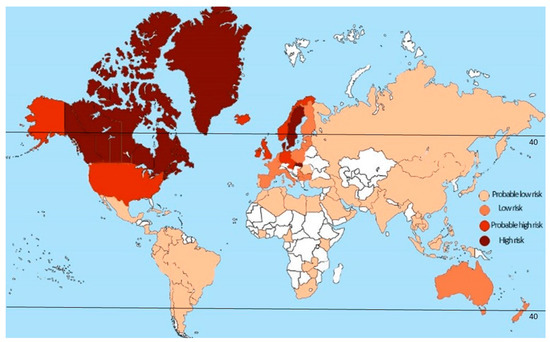
Figure 3. Global risk of development of MS. The prevalence of MS increases further from the equator in either hemisphere. Prevalence is higher in North America and Europe (291 and 232 per 100,000 respectively) and lowest in South America, Sub-Saharan Africa and East Asia, at 58 per 100,000 respectively [88]. It appears that highest incidence rate is registered among patients residing above the 42° latitude.There is limited direct evidence to explain the role of the latitude in pathophysiology of MS. The most plausible explanation of why more MS cases are diagnosed at higher latitude is less sunlight exposure. Specifically, lower vitD production in those with less sun exposure has been suggested as an explanation for the latitude-associated differences in MS risk [89]. It has been proposed that the intensity of ultraviolet B (UVB) radiation, which is essential for vitD synthesis in the skin [90], is lower at northern latitudes [91]. This is because UVB light is readily absorbed by the ozone layer [92]; therefore the increase in the ozone path length at higher latitudes results in decreased UVB light reaching the Earth [93]. This explains low vitD production in all individuals living at higher than 33° latitude [94]. Similarly, no vitD synthesis was shown in individuals living at 90° latitude and none was found during the winter at latitudes above 50° (Antarctica, most of Greenland and Alaska, northern parts of Canada, Russia and Europe) [95]. These authors demonstrated that little vitD production occurs outside of the summer in all of these northerly places. Also, a significant decrease in vitD synthesis was detected in individuals that relocated from southern regions.Interestingly, it appears that sunlight also has a direct immunosuppressive effect [96]. Using experimental autoimmune encephalomyelitis (EAE), an animal model of MS, Wang et al. have shown that UV radiation can ameliorate symptoms independent of vitD production [97]. Similar results were presented by Becklund et al. [98], where UV radiation inhibited inflammation and demyelination of the spinal cord. Also, UV radiation dramatically reduced CCL5 mRNA and protein synthesis as well as increased macrophage migration and IFN-γ production in the spinal cord. The authors suggest that UV radiation prevents the migration of inflammatory cells into the CNS by focal chemokine inhibition and a systemic increase of immunosuppressive IL-10.
vitD
Decreased serum levels of vitD in MS cases were demonstrated in 1994 by Nieves et al. [99]. Since then, the role of vitD in the pathogenesis of MS has been well established, where a number of studies provide strong evidence for correlation between low vitD level and the risk of MS [100,101,102,103].
Although a safe vitD level was not identified, it appears that 63.3–75 nM/L serum level of vitD is protective, as a lower risk of MS was found in these individuals [104,105].
This assumption is supported by the report made by the Institute of Medicine (IOM) Committee to Review Dietary Reference Intakes for vitD and Calcium in 2011 regarding the dietary requirements for vitD [106].
According to this report, a serum level of vitD below 50 nM/L could lead to serious health consequences. Also, according to the guidelines of the IOM and the Endocrine society, the “vitD sufficiency threshold” is 75 nM/L, which also correlates with stable serum parathyroid hormone levels [58].
Unfortunately, the majority of population around the world has levels of vitD below the threshold [58,107].T
he serum level of vitD also changes depending of the month of the year, being lower during the winter and increasing in the summer in temperate regions, suggesting links to sun exposure. In another study, seasonal changes in vitD were found in the serum from a large American cohort, peaking in late summer [108].
Interestingly, changes in vitD differ between sexes, where vitD levels are lower in woman when compared to men [109].
More so, summer and autumn levels of vitD in women were significantly lower as compared to those in men.
This could be related to the higher frequency of MS diagnosis in woman as compared to men [55,56,57].
Interestingly, vitD levels during pregnancy could be a risk factor for MS diagnosis in the child.
Studies have shown that the month of birth correlates with MS risk, with a significant increase in MS risk among individuals born in April and May as compared to those born in October and November [110,111].
This observation indicates the association between maternal sunlight exposure and the vitD status of the mother during pregnancy and vitD deficiency of the fetus.vitD replacement therapy has demonstrated an improvement in the mental health and decreased annual relapse rate in treated MS patients in several medium-sized clinical trials [112,113].
A large data met-analysis revealed a trend towards fewer relapses and improvement in expanded disability status scale EDSS for analyzed studies [114].
In addition to its well-documented role in calcium metabolism it is now clear that vitD also plays a significant role in immune signaling.
The receptor for the activated form of vitD (calcitriol), (nVDR) is present in many cell types including monocytes, macrophages and lymphocytes, and functions as a nuclear ligand binding domain, regulating transcription of many different genes.
The primary immune-modulatory effects of the calcitriol/nVDR complex are via modification of activation, differentiation and proliferation of immune cells.
Type I pro-inflammatory cytokines such as IFN-γ are downregulated and type II anti-inflammatory cytokines (such as IL-10) are upregulated.
The generation and activation of Treg and tolerogenic DC’s also occur in response to calcitriol signaling.
The overall effect of calcitriol signaling is a shift away from inflammatory immune responses. vitD also directly modulates the function of brain pericytes (which maintain the blood brain barrier, BBB) by downregulating inflammatory responses in these cells [115].
Clearly calcitriol-mediated immune modulation is in the opposite direction to that which characterizes inflammation in MS disease.
The production of vitD in response to sunlight exposure also clearly links to the latitude of habitation (which determines UVB exposure). These two risk factors are intrinsically tied together and provide an obvious reason for seasonal variation in MS relapses and the month of birth as an MS risk factor [116].
he overall effect of calcitriol signaling is a shift away from inflammatory immune responses. vitD also directly modulates the function of brain pericytes (which maintain the blood brain barrier, BBB) by downregulating inflammatory responses in these cells [115].
Clearly calcitriol-mediated immune modulation is in the opposite direction to that which characterizes inflammation in MS disease.
The production of vitD in response to sunlight exposure also clearly links to the latitude of habitation (which determines UVB exposure). These two risk factors are intrinsically tied together and provide an obvious reason for seasonal variation in MS relapses and the month of birth as an MS risk factor [116].
Lifestyle Factors
There have also been a number of studies linking lifestyle factors such as obesity, diet, changes in the gut microbiome, smoking, exposure to industrial chemicals such as organic solvents and an “urban” lifestyle with an increased risk of developing MS or of an increased risk of disease progression [117,118,119,120,121,122].
Much of the data for dietary and microbiome effects are inconsistent and there is little clear mechanistic explanation in many cases for how these factors are specifically linked to MS beyond the fact that poor diet, obesity, smoking and industrial chemical exposure are well recognized as inductive of a general pro-inflammatory state that could exacerbate other MS risk factors [123,124]
Viruses and Other Infecitous Agents
Environmental factors including viruses, where Epstein Barr Virus (EBV) infection and endogenous retrovirus reactivation have been connected to MS.
EBV is a gamma herpesvirus which has an almost ubiquitous prevalence in the adult population worldwide with more than 90% of adults demonstrating seroconversion to the virus.
It is spread by saliva and other secretions and primarily replicates in B cells, triggering their proliferation.
The accepted pathogenesis of EBV-induced disease is that those infected when young (prepubertal) do not show overt disease, whereas those infected as adolescents or adults will display infectious mononucleosis (IM or Glandular Fever), involving swollen lymph nodes, malaise and fever due to CD8 T cell responses.
The reason for this age-related difference in pathogenesis is not clear but is probably related to the fact that there is a general switch from more innate immune system driven to more adaptive immune system responses with age.
Like many herpesviruses, EBV establishes latency in its target cells. Several proteins produced by the virus during latency that manipulate the host cells’ life cycle can also trigger B cell lymphomas.
This effect is more marked in immunosuppressed patients such as those with AIDS [125,126].The epidemiological evidence for the involvement of EBV in MS centers around studies that have demonstrated that the risk of MS is very much higher in those individuals that have suffered from IM than those that have not (2–3-fold higher risk) [125]; conversely, the risk of MS for those who are not seropositive for EBV (admittedly a small cohort) is 15 times lower than those that have had the virus [126].
In addition, antibodies and T cells responding to lytic phase proteins of EBV, indicating recent infection or re-activation of EBV, are also associated with MS.
Like all viruses, EBV triggers Th1 cell-mediated immune responses and like many herpesviruses it also encodes proteins that manipulate the host’s immune response for the virus advantage.
These include proteins that inhibit the production of antiviral cytokines such as IFNα and IFNγ and proteins that interfere with HLA loading of viral antigens and proteins that inhibit apoptosis of immune cells [127].
It could be suggested that the type of antiviral immune response triggered by EBV correlates with that seen in MS pathogenesis.Exactly how EBV infection (which occurs in the peripheral circulation) translates into CNS immune dysfunction in MS is not clear; current theories include leakage of EBV-infected B cells across the blood brain barrier triggering a pro-inflammatory environment, cross activation of antibodies against EBV towards myelin antigens, or dysregulation of the immune system due to EBV infection and manipulation of B cells and monocytes causing stimulation of auto-reactive T cells [128,129].
Conversely, seropositivity to Human Cytomegalovirus (CMV) a betaherpesvirus that also has a near ubiquitous prevalence in the adult population conveys a protective effect on MS risk [130,131].
However, this theory is clouded by frequent reports of CMV recrudescence in MS patients receiving immunosuppressive therapy [132,133] and that EAE susceptibility in adult mice can be induced by Murine Cytomegalvirus [133].
This confusing picture of pathogenesis, similar to that of EBV is most probably explained by the hygiene hypothesis, whereby exposure to generic infections early in life enhances the development of an immunoregulatory system that decreases autoreactive T cell activity later in life.
Further evidence supporting this theory comes from studies of other pathogens that are normally acquired in childhood in poor hygiene environments such as Helicobacter pylori [134] and parasites such as Toxoplasma gondii [135] and Trichuris trichuria exposure, which also displays a negative association with MS.
Though in the case of helminth parasites, there may also be direct immunosuppressive effects of excretory proteins from the worms that are being explored as potential MS therapies [136].
Endogenous Retrovirus Expression
A further viral risk factor is that of endogenous retroviral expression. Endogenous retroviruses are repetitive genomic sequences that are present in most, if not all vertebrates.
They are thought to arise via infection of germ line cells via infectious retroviruses, as the lifecycle of retroviruses includes copying themselves into the host cells genome.
The ones in the human genome are no longer active as infectious viruses but they are expressed both as RNA and proteins, and the regulatory elements in the longterm repeats at either end of the viruses interact with a variety of cellular transcription factors.
The link between ERV expression and MS has been controversial; however, recent systematic reviews and meta-analysis of a large number of studies have clearly established that MS patients over-express RNA from the HERV-W ERV family when compared with healthy controls [137].
Other ERV families the expression of which has been associated with MS include: HERV-H, and in particular an SNP on one member of the HERV-H group, HERVFc1 on the X chromosome and HERV-K (particularly the polymorphic alleles HERV-K113 and HERV-K18 which are absent in some individuals) though the evidence for these is not as strong as for the HERV-W group.
Single reports have also reported a link between MS and HRES and HERV-15 elements [137].
In terms of a plausible molecular basis for HERV-W in MS pathogenesis, expression of env proteins from this group of ERVs induces pro-inflammatory cytokines (such as TNFα) and can replace the adjuvant (typically Freunds adjuvant) used in triggering EAE in mice.
Interestingly, EBV infection of B cells reliably triggers the expression of HERV-W loci by binding of the EBV gp350 protein. HERV-W expression is also increased in IM (EBV affected) patients, tying together these two risk factors in MS disease [138].
HERV-W is also constitutively expressed in the central nervous system and is crucial in placental function. The HERV-W member syncytin-1 is responsible for trophoblast fusion in the formation of the syncytia in human placentas [139].
It is feasible that immune responses against HERV-W, triggered by over-expression in EBV infection, could carry across the BBB into the CNS triggering local inflammation.
The expression of HERV-W in pregnancy also provides a potential link with the role of steroid hormones (the expression of which is altered in pregnancy).In a recent study, Manzari et al. showed an association of MS with a specific genetic variant of the nuclear antigen 2 (EBNA-2) [140].
These data provide strong evidence supporting the role of EBV in MS pathogenesis. Interestingly, EBNA2 directly interacts with the cellular DNA-binding protein RBPJ-κ (recombination signal-binding protein J kappa), a ubiquitous protein of the Notch signaling pathway that plays an important role in MS pathogenesis. RBPJ was suggested as a possible autoantigen in MS as it has been demonstrated to bind to CSF-derived IgG in some MS patients [141].
LMP1, another EBV protein, can induce expression of immunoglobulin kappa light chains [142], which are also consistently found in MS cases [143]. One multicentre study has shown the diagnostic value of these kappa-free light chains in CSF in diagnosis of MS [144].
Source:
University of Geneva
Media Contacts:
Doron Merkler – University of Geneva
Image Source:
The image is credited to UNIGE.
Original Research: Open access
“Brain-resident memory T cells generated early in life predispose to autoimmune disease in mice”.Doron Merkler et al.
Science Translational Medicine. doi:10.1126/scitranslmed.aav5519


