


 Extract (6-MSITC) in Healthy Older Adults")
: An In-Depth Exploration into its Thermogenic Role and Social Significance")


Amyloid fibrils are a type of self-assembled proteins/peptides that take on a stacked sheet-like formation.
Amyloid fibril aggregates are known to be a cause of several diseases–including Alzheimer’s–and therefore, it is of immense scientific interest to understand how these aggregates can be broken.
Some types of amyloid fibrils also play a role in regulation of gene expression in some organisms.
It is also thought that the fiber-like formats appearing in these aggregates act as scaffolds on which to cultivate biomaterials.
Therefore, a suitable technique for breakdown or “dissociation” of amyloid protein fibrils is critical from the perspective of medical treatment, modification of biological structures and functions, and even biomaterial engineering.
A collaborative group of Japanese scientists from the IR Free Electron Laser Research Center at Tokyo University of Science and The Institute of Scientific and Industrial Research at Osaka University, consisting of Dr Takayasu Kawasaki, Prof Koichi Tsukiyama, and Asst Prof Akinori Irizawa, has now shown that a far-infrared (FIR) free-electron laser (FEL), called FIR-FEL, can be used to break down amyloid protein aggregates, which is a testament to the power of interdisciplinary scientific research.
This study has been published recently in Scientific Reports.
Kawasaki states, “We wanted to demonstrate the applicability of strong free-electron lasers in the life sciences, and this interdisciplinary research has made this possible.”
Previous studies have investigated the dissociation of amyloid fibrils but with limited success and mixed results.
Because their dissociation in water is difficult, physical methods of dissociation have been explored in the past.
Lasers and electromagnetic radiation have been used for fabrication and structural/functional alteration of chemical and biological materials.
Among lasers, the FIR-FEL has been studied very sparsely, although it has high penetration power and is absorbed well by biological systems.
It is also used in tissue imaging, cancer diagnostics, and biophysics studies. Kawasaki explains, “Our study shows for the first time that FIR-FEL is also useful for breaking down the fibril aggregate structure of proteins.”
For their study, the researchers used the 5-residue peptide DFKNF as the model because the link between its fibrillation and pathogenesis is already established.
This peptide auto-assembles into a fibril sheet.
They found that FIR-FEL damaged the rigid β-sheet conformation (one of the few structures that proteins assume) of the 5-residue peptide by creating small holes on the peptide film.
The researchers found that FIR-FEL also disrupts the hydrogen bonds between adjacent β-sheets in the fibril and gives rise to free peptides.
This is referred to as dissociation.
Kawasaki and team then checked for conformational changes in the peptide fibril after irradiation with FIR-FEL, by analyzing the ratios of 4 types of secondary structures of peptides (α-helix, β-sheet, β-turn, and other).
They found that the proportion of the β-sheet conformation was drastically reduced, which suggests that the rigid sheet-like structure of the fibril was disrupted.
Kawasaki states that a previous study had also found mid-infrared (MIR)-FEL to be effective in this regard.
“We compared the effects of MIR-FEL with those of FIR-FEL,” says Kawasaki, “and we found that although MIR-FEL caused conformational changes in the fibril aggregates, it did not break down the fibrils as affectively as FIR-FEL did.”
Using scanning electron microscopy and dye staining techniques, the researchers also confirmed that FIR-FEL causes morphological changes in the fibrils.
Kawasaki says, “Because amyloid fibril peptides are involved in regulation of biological functions as well as pathologies, physical modification techniques (like FIR-FEL) could also be used to alter the biological functions of these macromolecules as needed.”

They found that the proportion of the β-sheet conformation was drastically reduced, which suggests that the rigid sheet-like structure of the fibril was disrupted. The image is in the public domain.
As FIR-FEL is more effective than MIR-FEL, FIR-FEL can be used to destroy amyloid fibrils deep inside tissues, as in the case of Alzheimer’s disease, whereas MIR-FEL can be used for removing dermal amyloids on the surface of the skin.
Also, because fibril proteins act as scaffolds for biocompatible materials, FIR-FEL could be used in biomaterial engineering in regenerative medicine or Nano carrier drug-delivery systems.
To conclude, Kawasaki eloquently states, “For the first time in the world, we have found that a rigid aggregate of amyloid fibrils can be effectively broken down using a free-electron laser in the terahertz region (wavelength 50-100 micrometers).
Our next step would be to understand how FIR-FEL affects different types of peptide fibrils.
Our research can fuel the development of novel treatments for intractable diseases such as Alzheimer’s. It could also aid the development of new methods for manipulating the structure of biocompatible materials.”
FIR-FEL
FIR-FEL developed at The Institute of Scientific and Industrial Research, Osaka University was used in this research (Fig. 1). The FIR-FEL can generate intense pulsed laser beam, and the features are briefly summarized as follows:20 (1) oscillation wavelengths are 50–100 μm (A). In this study, we prepared four different wavelengths, 60, 74, 80, and 97 μm for the irradiation experiments. The half width was about 5 μm in each spectrum. (2) Time structure is composed of micro- and macro-pulses (B): a micro-pulse has a duration of 10 pico-second (ps), and about one hundred micro-pulses are bunched in one macro-pulse with a 4 μs duration. The FEL oscillates at 5 Hz throughout the experiment. (3) Typical pulse energy used was from 0.5 to 1.5 mJ per macro-pulse (C).
MIR-FEL
Detailed oscillation system is described elsewhere21. In brief, mid-infrared (MIR)-FEL at Tokyo University of Science was operated at the mid-infrared region (5.0–10 μm: 1,000–2,000 cm−1), and the time structure is composed of macro- and micro-pulses, and a repetition rate of macro-pulse is 5 Hz similarly with the case of FIR-FEL, although micro-pulse has a duration of 1 to 2 ps and the interval of the consecutive micro-pulses is 350 ps. The pulse energy per a macro-pulse at around 6 μm was ca. 10 mJ.
Preparation of peptide fibril
The powder of DFNKF was dissolved in DMSO (100 mg/ml) for a stock solution22. This solution was diluted by ten times using tris-buffer (20 mM, pH7.5) containing sodium chloride (10 mM) and incubated at 37 °C for several days to produce the fibril. The resulted peptide fibril solution (20 μL) was spread on a stain less steel base to produce a thin film (thickness: ca. 0.1 mm; diameter: ca. 5 mm) for infrared microscopy analysis and on a glass slide base for scanning-electron microscopy observation and Congo-red staining as describe below. After drying under atmosphere for 6 hours, the peptide fibril was irradiated by FIR-FEL or MIR-FEL. The FEL beam was focused on the sample using a parabolic reflector for FIR-FEL or BaF2 lens for MIR-FEL. In both cases, the irradiation direction was vertical against the sample surface.
Terahertz spectroscopy
We used FT/IR-6700FV (Jasco Co., Tokyo, Japan) for measurement of infrared spectrum of peptide at terahertz region. The peptide film was prepared on a stain less steel base as described above, and the measurement was performed in vacuo by reflection mode. As for preparation of the film of pre-fibril, the stock solution of the peptide in DMSO was diluted by the tris-buffer and was immediately dropped onto the stain less steel base at room temperature. The spectrum was recorded with 512 scans.
Infrared microscopy
The IR spectral measurement was performed using IRT-7000 infrared microscope (Jasco Co, Tokyo, Japan) and FT/IR-6100 spectrometer (Jasco Co., Tokyo, Japan). The infrared spectra were recorded by a reflection mode from several areas on the dry surface of the peptide film with 64 scans and 4 cm−1 resolution, and the morphology of the peptide film was observed by 16x Cassegrain lens. The obtained spectral data were transformed into text files, and those absorption intensities at a range of wavenumbers from 1000 to 4000 cm−1 were averaged followed by re-converting to infrared spectral trace.
For protein secondary structure analysis, we used IR-SSE analytical software (Jasco Co., Tokyo, Japan). In this program, a calibration curve was prepared prior to multicomponent analysis (Partial Least Squares quantification model) based on the secondary-structural data of 17 proteins, and it was saved as a standard data file23. The amide I band was deconvoluted into major four bands: α-helix (1650–55 cm−1), β-sheet (1625–40 cm−1), β-turn (1655–75 cm−1), and non-ordered (other) conformation (1645–50 cm−1). Proportions of secondary structures were calculated using peak intensities at those amide-I bands in the averaged IR spectrum.
Scanning-electron microscopy (SEM)
We used FE-SEM Supra40 scanning electron microscope (Carl Zeiss). After the peptide fibril was added on a glass slide base and irradiated by the FEL as described above, the slide base was fixed on a sample holder by using conductive copper tape, and the surface of the fibrils was observed using the acceleration voltage at 5.0 kV.
Congo-red staining
The stain solution was prepared by dissolving powder of Congo-red in phosphate-buffered saline to be 0.2 mM concentration. This solution (10 μL) was added on the peptide fibrils on a slide base, and the mixture was incubated for at least 10 min at room temperature. After drying, the surface was observed by using a polarized light microscope MVX 10 (Olympus, Tokyo).
Reduction of β-sheet conformation by the terahertz radiation
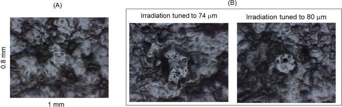
Both of irradiations gave visible damage on the peptide film after 30 min irradiation (~9000 pulses) with ca. 1 mJ energy per a macro-pulse: small holes were obviously observed (Fig. 3B) compared to the fibril before irradiation (Fig. 3A).
The sizes of these spots were ~200–300 μm in diameter that corresponds to the focused beam diameter, in which a clear circle was seen in case of irradiation at 80 μm compared to that at 74 μm.
The infrared absorption spectra, recorded from several areas as shown by yellow squares in the surrounding debris (Fig. 4A), were averaged and compared before and after the irradiation (Fig. 4B, see Supplementary Materials). Doublet structure of the amide I peaks from 1600 to 1700 cm−1 are common spectral features of the peptide in the mid-infrared region (left panel). Reches et al.22 attributed the amide I peak at lower wavenumber (~1645 cm−1) to the C=O stretching vibrational mode of amide bonds in β-sheet stacked structure, because (i) the peak intensity at ~1645 cm−1 is weaker than that at higher wavenumber (~1670 cm−1) before fibrillation (Pre-fibril) and (ii) the former intensity increased after fibrillation (Non-irradiation fibril).
It should be noted that the peak intensity at higher wavenumber increased after irradiation at 74 and 80 μm, and the double peaks were slightly shifted to higher wavenumbers (plus ca. 10 cm−1) after those irradiations, while amide II band (at ~1550 cm−1) and a tiny peak at 1500 cm−1 little moved.
These spectra show that conformations regulated by the amide backbone in the fibril state were changed by the irradiation, and the blue shift of the amide I bands and an increase of peak intensity at higher wavenumber (~1670 cm−1) may indicate that rigid conformation was raveled out to the non-aggregate form.
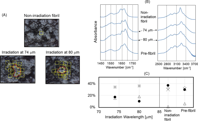
In the near infrared region (right panel), the O–H stretching vibrational band was observed at around 3250 cm−1.
The sharp peak was observed for the fibril state (top), and the broadened band with shoulder at 3300–3400 cm−1 was characteristic for the pre-fibril state (bottom). Interestingly, irradiations at 74 and 80 μm to the fibril state afforded the similar spectra as that of the pre-fibril state: the peak at 3250 cm−1 was broadened and a shoulder peak at ~3300 cm−1 was clearly seen.
These near-infrared spectra support FIR-FEL driven conformational change in the fibril structure at specific far-infrared wavelengths.
Together with the spectral change in the amide I region, the FEL irradiation may disrupt hydrogen bonds between β-sheets in the fibril state and produce non-aggregated free peptide chains.
To estimate the conformational change of the peptide fibril in more detail, the ratio of four types of secondary conformations (α-helix, β-sheet, β-turn, and other) were derived through the spectral deconvolution of the amide I band (Fig. 4C, see also Supplementary Materials).
For non-irradiation fibril (right panel), the content of β-sheet (black circle) was ~40%, those of β-turn (cross) and other conformations (diamond) were ~30% each, and α-helix (triangle) was hardly detected. For pre-fibril state, α-helix was recognized (~5%), and the β-sheet content was slightly lower than that of non-irradiation fibril state.
In contrast, both irradiations at 74 and 80 μm substantially reduced the amount of β-sheet down to about 10‒20% and increased that of α-helix to near 20% (left panel). Interestingly, the amount of β-sheet was lower and that of α-helix was higher in both cases than those in the pre-fibril state.
In addition, it seems that the irradiation at 80 μm reduces the β-sheet conformation in the fibril more remarkably than that at 74 μm.
Source:
Tokyo University of Science
Media Contacts:
Tsutomu Shimizu – Tokyo University of Science
Image Source:
The image is in the public domain.
Original Research: Open access
“Dissolution of a fibrous peptide by terahertz free electron laser”. Takayasu Kawasaki, Koichi Tsukiyama & Akinori Irizawa.
Scientific Reports. doi:10.1038/s41598-019-47011-z


