


 Extract (6-MSITC) in Healthy Older Adults")
: An In-Depth Exploration into its Thermogenic Role and Social Significance")


Getting a close look at the prostate is critical for detecting cancer, but its rather intimate positioning (just in front of the rectum) makes it difficult to image.
Now, professor and chair of radiology Sanjiv “Sam” Gambhir, MD, Ph.D., thinks he has a solution: a newly devised hybrid camera.
Traditionally, prostate cancer is detected via prostate cancer-associated blood biomarkers, such as prostate-specific antigen (PSA).
Doctors often also use ultrasound or magnetic resonance imaging (MRI) to look for physical changes in prostate tissue.
Newer techniques that harness positron emission tomography (PET) scans can even capture molecular detail, but those tactics are relatively more expensive and use radiation, said Gambhir.
“But the problem is that cancer within the prostate quite often doesn’t lead to any anatomical changes until it’s quite large or has spread beyond the capsule of the prostate into the lymph nodes around it,” said Gambhir. “So for decades we’ve been looking for ways to analyze and image the prostate with greater detail to detect changes earlier on, safely, and at relatively low cost.”
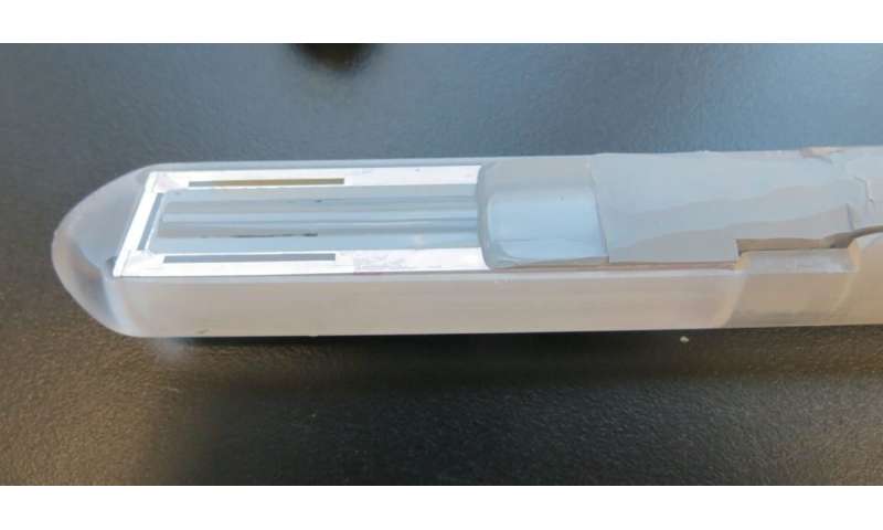
Gambhir’s new technology, dubbed the transrectal ultrasound and photoacoustic device, or TRUSPA, marries ultrasound and photoacoustic imaging techniques to simultaneously produce a picture showing the anatomy of the prostate, functional details about the gland and molecular information that can help flag cancerous tissue.
In a proof of principle study, Gambhir and a team of scientists across Stanford, including biologists, engineers and doctors, have demonstrated the value of the instrument in about 20 patients.
A paper describing the technology and results published in Science Translational Medicine. Gambhir is the senior author of the study and Sri-Rajasekhar Kothapalli, Ph.D., is the lead author.
The TRUSPA platform combines ultrasound (gray) and photoacoustic (red) imaging to visualize the prostate in real time. Credit: S.R. Kothapalli et al., Science Translational Medicine (2019)
Since ultrasound is already used widely by urologists and generally in human imaging, Gambhir opted to start with that as the foundation of TRUSPA.
Typically, if a biomarker such as PSA is elevated in a patient’s blood, doctors then turn to a combination of ultrasound and biopsy, during which they use a needle to take about 20 samples from different regions of the prostate.
The technique is rooted in a poke-and-hope theory, as in, hopefully you’re sampling the part of the prostate that contains the cancer tissue. But it’s not guaranteed.
TRUSPA takes a different approach, which incorporates an imaging agent that cancer cells readily take up—more so than regular tissue.
Then, through photoacoustic molecular imaging (which monitors the absorption of light waves to help characterize tissue type) doctors can see where the cancer cells are located in the prostate.
The presence of the imaging agent in tumor tissue changes the way that the light gets absorbed and ultrasound waves are sent back to the device, making it into a sort of flag for cancerous tissue.
“We opted for an imaging agent that was not specific to prostate cancer, but rather to cancerous tissues for our proof of principle,” said Gambhir.
That imaging agent was already FDA approved, making it an easy starting point. “But the idea moving forward is to heighten precision using a molecularly-targeted photoacoustic molecular imaging agent that binds specifically to prostate cancer cells.”
In the pilot study, the scientists used the device in 20 individuals who had been diagnosed with prostate cancer, looking to see whether or not their device could likewise detect the disease.
“Not only were we able to better understand TRUSPA imaging limitations in the patients, we also were likely seeing tumors that would have otherwise been invisible to conventional prostate ultrasound,” said Gambhir.
Credit: Stanford University
In one patient, they were even able to differentiate between malignant and non-malignant cancer tissue, which was later confirmed upon further molecular analysis when the diseased prostate was removed from the patient.
Gambhir cautions that this is still a pilot, and that the team need to test the imaging system much more before we conclude that TRUSPA can make these sorts of differentiations broadly. But it is a promising start, he said.
With clear evidence that the concept and technology can be made to work in humans, now Gambhir and his team are continuing to improve the device, it’s spatial resolution, the molecular photoacoutic imaging agents specific for prostate cancer and more to enhance the accuracy and sensitivity of tumor detection.
“We’re now starting to explore TRUSPA for detecting ovarian cancer, thyroid cancer and skin cancer, too,” said Gambhir.
Butrus (Pierre) Khuri-Yakub, Ph.D., professor of electrical engineering at Stanford, Geoffrey Sonn, MD, assistant professor of urology, Joseph Liao, MD, associate professor of urology and James Brooks, MD, professor of urology, also contributed to the project.
Molecular imaging is quickly gaining importance in both biomedical research and clinical diagnostics. During the early phases of cancer development, biochemical alterations of cell metabolism occur before the formation of detectable tumour masses.
Current molecular imaging techniques, targeted to the study of molecular kinetics, employ molecular tracers capable of detecting cancer lesions with both high sensitivity and specificity while also providing essential information for both prognosis and therapy. On the contrary, crucial information is provided by histopathological examination and ancillary techniques such as immunohistochemistry.
Indeed, in situ analysis of cancer biomarkers by immunohistochemistry provides prognostic and predictive information related to the metabolic characteristics of the tumour mass. Therefore, a multidisciplinary approach based on the collaboration between diagnostic imaging and anatomic pathology could improve the management of oncological patients, further supporting the current goal of a completely personalized medicine approach in oncology.
In detail, this new approach will allow clinical doctors to investigate the living body to identify disease, monitor progression, or treat medical conditions at a molecular level [1]. In this review, we discuss the possible alliance between diagnostic imaging and pathology with a focus on teamwork between nuclear medicine and anatomic pathology.
2. Diagnostic Imaging and Anatomic Pathology: An Alliance for the Diagnosis of Oncological Patients
The use of diagnostic imaging has increased steadily over the past decade, dramatically contributing to improved management of oncological patients. Indeed, high-resolution images provided by computed tomography (CT), magnetic resonance imaging (MRI), and ultrasound (US) allow early detection of a vast number of tumour masses. In this context, combined imaging based on positron-emission tomography- (PET-) CT plays a central role in the diagnosis, staging, and posttreatment follow-up of patients with neoplastic diseases [2], and several studies have shown the increase in diagnostic value resulting from combined PET-CT. For instance, Lardinois et al. reported for the first time the advantage of PET-CT over other techniques for TNM staging [3]. In addition, recent studies have shown that PET-CT is the best imaging modality for tumour staging and specifically that it is superior both to PET and to PET and CT performed separately [4]. Combined PET-CT acquisition can therefore improve the diagnostic value of the examination and implements the concept of “one-stop shop” recently introduced in modern clinical medicine, which can also be extended to more recent PET-MR developments (see below) [5]. The possibility of performing TNM staging and formulating a full diagnosis with a single examination also drastically reduces the time commonly required to perform all diagnostic examinations separately [2].
While radiography, ultrasonography, and CT are the main imaging techniques employed in cancer diagnosis, MRI is an emerging imaging diagnosis method that, while well established in the clinical practice, is in continuous development. Specifically, MRI can produce three-dimensional, multimodal images in a noninvasive way, without the use of ionizing radiation and with exceptional spatial and contrast resolution, allowing unprecedented accuracy in tumour investigation [6]. The novelty of MRI analysis is focused mainly on improving the anatomical resolution and on the advent of functional as well as molecular approaches [7]. Currently, available MRI techniques allow both structural assessment and evaluation of different physiopathological processes of the tumour microenvironment [7, 8]. Also, structural and functional MRI results in a more comprehensive evaluation of the extension and activity of neoplastic diseases [9]. Of note, a correct oncologic status evaluation allows for the establishment of better therapeutic strategies, with a favourable impact on prognosis as well as survival. In addition, the use of perfusion, spectroscopy, and diffusion MRI modalities can provide essential data about angiogenesis, cell metabolism, and cellularity of cancer lesions [10–12]. Still, histopathological studies are essential to validate the results of PET-CT and MRI exams [13]. Analysis of biopsies or surgical specimens still represents the gold standard for the diagnosis and full characterization of neoplastic lesions [14]. Moreover, the introduction of novel and high-performance molecular biology techniques on paraffin sections allows to compare metabolic data of molecular imaging (i.e., uptake of FDG or radiolabelled antibodies) with the in situ localization of molecules involved in cancer pathways. As concerns the relationship between MRI and histopathological data, breast cancer in particular offers important perspectives. Indeed, several studies have focused on the identification of MRI features that can predict the molecular patterns of breast cancer such as ER, PR, Ki67, and HER2 status [15]. In this context, Ouk et al. reported that triple-negative breast cancer (TNBC) was larger and better defined and had more necrotic tissue compared to other cancers and that necrosis yielded high T2-weighted signal intensity and apparent diffusion coefficient (ADC) values [16]. Uematsu et al. found similar MR features for TN cancers, including high intratumoural signal intensity on T2-weighted MR imaging and intratumoural necrosis [17]. Bae et al. reported that, in MRI, androgen-receptor-positive TN cancers were more likely to be associated with nonmass types and a higher incidence of irregular and spiculated lesions than androgen-receptor-negative TN cancers [18]. Also, a recent study demonstrated a correlation between HER2 overexpressed cancers and persistent enhancement in the delayed phase on MRI, suggesting that MRI can be potentially useful in the diagnosis and subtyping of breast cancer. The information obtained by immunohistochemical analysis can generate greater concordance when compared with molecular imaging techniques such as PET and SPECT (single-photon-emission computerized tomography). In this scenario, nuclear medicine and anatomic pathology share several common interests. They constitute two fundamental approaches for the establishment of diagnosis, clinical monitoring, prognosis, and therapy response. Both disciplines are similarly interested in finding anatomic lesions and in deepening knowledge about their nature at a cellular and molecular level [19, 20]. More than ever before, nuclear imaging is playing an integral role during both initial investigations and follow-up of patients with acute and chronic illnesses. It comprises several techniques, such as PET and SPECT, that use specific radiopharmaceuticals to image certain molecular interactions belonging to biological processes in vivo and are therefore able to deliver information about biomarker expression, tumour burden, tumour metabolism, receptor status, and proliferation. In particular, the spatial resolution of PET-CT offers exceptional insights into the human body that enable physicians to identify and characterize tumoural lesions in the early stages of their development [21–23]. It is important to note that, in some cases, it is possible to use the same antibodies to detect target molecules both in vivo by immuno-PET analysis and ex vivo by immunohistochemistry [24]. The treatment of breast and prostate cancers is based on immunohistochemical analysis of well-known prognostic markers such as ER, PR, Ki67, cerb2 (Figure 1), or PSA [25, 26]. Nevertheless, cancer cells frequently undergo selection during pharmacological treatment, hence possibly changing their dominant molecular phenotype and leading to drug resistance. Even though heterogeneous temporal evolution of cancer cells can be evaluated through sequential biopsies, this would imply the necessity of repeated invasive procedures. To reduce the number of biopsies and simultaneously increase the patient’s quality of life, PET radiolabelled molecules directed against markers which are prognostic and predictive in cancer (i.e., radiolabelled anti-cerb2 antibodies [27] or radiolabelled PSMA inhibitors [28]) have been developed. In the last years, several studies identify some significant correlations among histological parameters, immunohistochemical data, and 18F‐FDG uptake (PET‐MRI analysis) in patients affected by head or neck squamous-cell carcinoma [29, 30]. All at once, these data can lay the foundation for the development of new in vivo analysis capable of predicting prognosis and therapy response early without the support of histological analysis. Specifically, histological samples offer the possibility to characterize cancer lesions from a genetic point of view, by studying chromosomal alterations like gene amplification or DNA fragment translocation [31], and at an ultrastructural and atomic level, thanks to the performance of scanning and transmission electron microscopes and their ancillary technologies (such as energy-dispersive X-ray microanalysis and immunogold labelling) [32–35]. These new approaches may be considered a further element capable of “linking” molecular imaging and anatomic pathology. A great proof of nuclear medicine and pathology collaboration is the characterization of breast microcalcification. Indeed, recent studies, focused on exploring the clinical and biological significance of breast microcalcifications, demonstrated that their elemental composition and histological aspect were related to patients’ prognosis [36, 37]. In addition, in these studies, it was shown that microcalcifications made of hydroxyapatite are actively produced by cancer osteoblast-like cells (OLCs) in a process similar to what occurs during physiological bone matrix formation [38–40]. On the basis of this evidence, molecular imaging could be used to discriminate cancer from any other type of lesion by studying the metabolic activity of breast tissues associated with the presence of microcalcifications as shown by Ahrens et al. in a mouse model [41].

Evaluation of in situ breast cancer biomarkers. Image showing (a) Ki67 nuclear expression in an infiltrating breast cancer, (b) expression of ER in an infiltrating breast cancer, (c) PR expression in an infiltrating breast cancer, and (d) HER2 expression (score 3) in an infiltrating breast cancer.
The affinity between nuclear medicine- and anatomic pathology-based diagnostics can therefore lay the basis for the creation of a synergic framework at the service of personalized medicine.
3. Digital Pathology
Since the 1980s, radiology has been revolutionized by the introduction of digital imaging, with a resulting improvement in quality and safety of reporting as well as disrupting innovation in the analysis and manipulation of radiological images [42]. Also, the recent introduction of digital pathology and whole slide imaging (WSI) (i.e., the complete digitization of slides) in anatomic pathology is transforming the practice of histodiagnostics [43]. During the last few decades, the introduction of digital cameras producing still images and microscope-mounted video cameras that allow live examination of slides has radically changed optical pathology [44]. In addition, approximately a decade ago, further improvements of these techniques resulted from the creation of digital slide scanners. These still or dynamic images can be transferred to remote sites to be assessed by another pathologist—that is, what is commonly called telepathology [45, 46]. This has facilitated applications such as teleconsultation and frozen section diagnosis [47].
WSI produces high-resolution digital images and involves relatively high-speed digitization of glass slides of different samples (e.g., tissue sections, smears, and thin-layer cytology), scanning them at multiple magnifications and focal planes (x, y, and z axes) [48]. Compared to static and live digital images, WSI is generally more beneficial. For educational and training purposes, WSI is more interactive and easy to share and provides access to the entire slide to help answer “on-the-spot” clinical questions at tumour boards. One can also generate teaching sets (virtual slide boxes) that can include a wide case range as well as rare cases that do not fade, break, or disappear. The digital imaging process includes four key steps: (a) image acquisition or capture; (b) storage and management, manipulation, annotation, and editing; and (c) viewing, displaying, or sharing of images. From a clinical point of view, digital images can be used for primary diagnosis, second opinions, telepathology, quality assurance (e.g., re-review and proficiency testing), archiving and sharing, training, education and conferencing, image analysis, research and publications, and marketing and business purposes, as well as tracking. Unfortunately, to date, the operational phases of digital pathology have not yet been standardized. Before digital slides are widely used for routine clinical analysis, standard procedures are needed and the entire imaging process is validated through large-cohort studies. However, in 2016, García-Rojo [49] released the first document with the guidelines and position papers from the Canadian Association of Pathologists, the College of American Pathologists, the American Telemedicine Association, the Digital Pathology Association, the Food and Drug Administration, the Centers for Medicare and Medicaid Services, the Centers for Disease Control and Prevention, the Society of Toxicologic Pathology, the European Commission, the Spanish Society of Anatomic Pathology, the Royal College of Pathologists, and the Royal College of Pathologists of Australasia.
Still, automated image analysis will enhance diagnostic efficacy in histopathology. Because inspection of WSI is probably slightly more time-consuming than that of conventional slides [50], the creation of software packages for detecting regions of interest will be advantageous and speed up the workflow, especially if those areas of interest could be outlined before the pathologist sees the image. To this end, grid computing would probably be needed to be able to apply several algorithms to WSI [51]. Still, software for computerized quantification of immunohistochemically stained WSI to improve the objective assessment of the immunoreactivity is already available from several scanner vendors. Such software estimates colour intensity relative to control cells and has Food and Drug Administration regulatory approval in the United States for the quantification of nuclear markers such as estrogen receptor (ER) or cell membrane markers such as HER2. Notably, the treatment of breast cancer patients is often based on immunohistochemical analysis of ER, progesterone receptor (PgR), Ki67, and HER2 [42, 52] (Figure 1).
Despite that new technologies open the possibility to detect digital spatial profiling of proteins and RNA in fixed tissue [53], currently diagnostic immunohistochemical analysis cannot capture the known spatial heterogeneity of breast cancer defined at the intratumoural, intrametastatic, and intermetastatic levels. Thus, the quantification of immunohistochemical data can be used to design new molecular diagnostic imaging or therapeutic protocols (PET- or SPECT-CT) that improve the patient’s quality of life by (a) reducing the number of biopsies, (b) increasing the accuracy of the diagnosis, and (c) identifying in “real time” the molecular transformation of cancerous cells. In particular, the use of radiolabelled molecules capable of quantifying the expression of ER (i.e., 16α–18F-fluoro-17β-estradiol) or cerb2 (i.e., radiolabelled anti-HER2 antibodies) by PET-CT could represent an alternative and reliable method for the management of breast cancer patients [54]. Our recent studies highlighted the possible role of 99mTc sestamibi SPECT as a possible tool for early detection of breast cancer lesions with high bone metastatic potential (Figures (Figures22 and and3)3) [40, 55]. In particular, we showed a putative correlation between 99mTc sestamibi uptake and aggressiveness of the tumours. High sestamibi uptake was detected in both triple-negative breast cancer lesions (Figure 2) and poorly differentiated infiltrating breast carcinomas forming lymph node (Figure 3) and bone metastasis.

Figure 3
99mTc sestamibi SPECT analysis and histological evaluation in a breast cancer patient. (a) Image showing sestamibi uptake in a breast cancer patients. (b) Histological evaluation (H&E) of the breast biopsy of the patient subjected to 99mTc sestamibi SPECT. (c) Sentinel lymph node with macrometastasis. Immunohistochemical analysis of lymph nodes displaying triple-positive phenotypes of breast cancer cells: (d) ER positive; (e) PR positive; (f) HER2 score 3.

99mTc sestamibi SPECT analysis and histological evaluation in a breast cancer patient. (a) Image showing sestamibi uptake in a breast cancer patients. (b) Histological evaluation (H&E) of the breast biopsy of the patient subjected to 99mTc sestamibi SPECT. (c) Sentinel lymph node with macrometastasis. Immunohistochemical analysis of lymph nodes displaying triple-positive phenotypes of breast cancer cells: (d) ER positive; (e) PR positive; (f) HER2 score 3.
Similarly, the search for new biomarkers of prostate cancer is constantly advancing. The most important radiotracer used for diagnosis and staging of prostate cancer is choline (11C-choline and 18F-choline) (Figure 4) [56]. Currently, choline PET and PET-CT techniques deliver high sensitivity and specificity for the diagnosis of locoregional and distant metastases in prostate cancer patients with recurrence of disease [56]. In our experience, a high 18F-choline signal is significantly associated with PSA plasma levels, histological grading, and in situ PSA expression (Figure 4). The comparative expression of PSMA on prostate biopsies analyzed through digital pathology and molecular imaging techniques can further improve the accuracy of nuclear medicine analysis by providing elements for both identification of new PSMA inhibitors and comprehension of biological mechanisms related to PSMA expression in prostate lesions. In this context, Bernacki et al. demonstrated that anti-PSMA immunohistochemical analysis is a promising marker for the identification of metastatic prostate carcinomas in surgical specimens [57].

18F-choline PET analysis and histological evaluation in a prostate cancer patient. Images showing (a, b) the uptake of 18F-choline in a prostate cancer lesion, (c) PSA expression in a prostate cancer biopsy, and (d) ck34be12 expression in a prostate cancer.
More information: Sri-Rajasekhar Kothapalli et al. Simultaneous transrectal ultrasound and photoacoustic human prostate imaging, Science Translational Medicine (2019). DOI: 10.1126/scitranslmed.aav2169
Journal information: Science Translational Medicine
Provided by Stanford University


