


 Extract (6-MSITC) in Healthy Older Adults")
: An In-Depth Exploration into its Thermogenic Role and Social Significance")


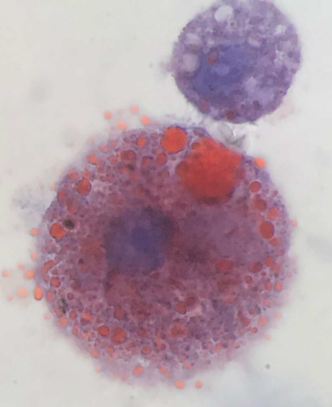
Doctors have identified a previously unrecognized characteristic of the vaping-related respiratory illness that has been emerging in clusters across the U.S. in recent months. Within the lungs of these patients are large immune cells containing numerous oily droplets, called lipid-laden macrophages.
The finding may allow doctors to definitively diagnose the nascent syndrome more quickly and provide the right treatment sooner.
It could also provide clues into the causes of the new and mysterious condition. Investigators at University of Utah Health reported the findings in a letter published in the New England Journal of Medicine on Sept. 6.
“While it is too soon to be sure, these lipid-laden macrophages may turn out to be useful to confirm or rule out this disease,” said the study’s senior author Scott Aberegg, M.D., a critical care pulmonologist at U of U Health.
“They may also be helpful in understanding what is causing this illness,” Aberegg added.
Lung scans from patients with vaping illness look like a serious viral or bacterial pneumonia, but those tests come back negative.
Instead, diagnosis has been based on exclusion of known causes of similar respiratory illnesses combined with knowing the patient has a history of vaping.
U of U Health investigators identified the lipid-laden macrophages in six out of six cases seen at University of Utah Hospital in Salt Lake City by the time of submitting the results for publication.
The cells were found in samples after performing a procedure called bronchoalveolar lavage where fluid is squirted into a small section of the lungs and then collected for examination.
Macrophages are a type of cell from the immune system that gather at sites of infection and perform the role of cleaning up debris.
Staining them with a dye called Oil-red-O highlighted the oily droplets littered throughout these cells.
“These cells are very distinctive, and we don’t often see them.
That made everybody start to think carefully about why they were there.
Are they scavenging debris in the lungs that was introduced through vaping?” Aberegg said.
Doctors at the U had the test performed on the first vaping patient treated at U of U Health in July 2019, after the referring doctor had suggested the patient could have lipoid pneumonia.
The condition is diagnosed by screening for lipid-laden macrophages.
After finding the marker in this patient, doctors performed the same test in subsequent patients suspected to have the vaping illness, and all were positive.
Since submitting their findings for publication, the number of vaping illness cases with lipid-laden macrophages has risen to ten of ten patients examined, with new cases arriving weekly.
The question remains as to whether the vaping respiratory illness is a type of lipoid pneumonia.
Despite similarities, there are also differences.
Unlike the vaping illness, classic lipoid pneumonia is typically seen in older individuals, typically caused by accidentally breathing in oil-based laxatives.
Classic lipoid pneumonia also presents differently on x-rays of the lungs.
Additional testing will need to be done to determine whether the vaping illness can be categorized as a new kind of lipoid pneumonia.
“We need to determine if these cells are specific for the illness or whether they are also seen in vaping patients who are not ill and don’t have symptoms.
If they are only seen in patients who get sick, we can begin to make some connections between what we’re seeing in the lipid laden macrophages and whatever components of the vaping oils may be causing this syndrome” Aberegg said.
Macrophages are found in most tissues including the lungs and airways where they play an important role in immune surveillance.
They are responsible for the phagocytosis and endocytosis of dying cells and pathogens.
Sputum macrophages from asthmatic subjects exhibit a phagocytic response that is inversely correlated with sputum eosinophilia and positively correlated with FEV1 and PC20FEV1 [1].
This suggests that decreased phagocytosis relates to airway inflammation and asthma severity.
In contrast, sputum-derived macrophages from asthmatic patients with no sputum eosinophilia display impaired efferocytosis of apoptotic bronchial epithelial cells [2].
In addition, a reduced phagocytic response to Staphylococcus aureus [3] in macrophages from children with severe asthma has been related to the presence of oxidative stress as assessed by glutathione oxidation [4].
Alveolar macrophages (AMs) that become laden with lipid (predominantly cholesterol) are termed pulmonary foam cells or lipid-laden alveolar macrophages (LLAMs).
These macrophages are more likely to undergo the process of programmed cell death and exhibit impaired phagocytosis [5], which may play an important pathogenic role in asthma.
Furthermore, markers of lipid peroxidation measured in plasma are increased in asthma subjects [6] whilst erythrocytes and platelets exhibit alterations in membrane fatty acid composition [7].
A high fat content diet and the administration of a lipid emulsion in rats has been associated with the formation of LLAMs, leading to the view that the lungs may be a potential route of lipid excretion.
However, it remains unclear whether such cells are derived from AMs ingesting lipids in the alveoli or from lipid-laden blood monocytes that migrate to the alveoli [8].
Lipid accumulation in alveolar macrophages appears to occur primarily through the phagocytosis of external lipoproteins.
This process may be secondary to endogenous sources, such as lipoid pneumonia; where the presence of bronchial obstruction, chronic lung infection or a lipid storage disorder results in the accumulation of LLAMs; or an exogenous source, for example following aspiration or inhalation of lipid [9].
There are other factors which have been associated with an increase in LLAM formation.
These include gastroesophageal reflux disease (GORD) [10], [11], hypoxia [12] and iatrogenic causes, whereby drugs such as amiodarone, fluoxetine, and gentamicin containing a cationic amphiphilic structure can induce cellular phospholiposis through a dose-dependent process involving the inhibition of lysosomal phospholipase activity and accumulation of lamellar bodies [13], [14].
In addition, macrophages have been shown to internalise and degrade surfactant lipids and surfactant protein A (SP-A) in vitro, suggesting a role for AMs in surfactant clearance [15].
Other factors such as obesity and cholesterol may also influence lipid-laden macrophage formation, but their role has not yet been established.
The aim of this study was to explore the relationship between the presence of LLAMs, chronic cough and asthma severity in a cohort of patients with mild/moderate and severe asthma. In addition, we determined whether obesity, the presence of GORD, lung inflammation or the degree of airflow obstruction influenced the accumulation of intracellular lipids.
More information: The research publishes on Sept. 6 as a letter to the editor at the New England Journal of Medicine, titled, “Pulmonary disease and lipid-laden macrophages associated with e-cigarette use”.
Journal information: New England Journal of Medicine
Provided by University of Utah


