


 Extract (6-MSITC) in Healthy Older Adults")
: An In-Depth Exploration into its Thermogenic Role and Social Significance")


Cancer treatment could be dramatically improved by an invention at the University of Waterloo to precisely locate the edges of tumors during surgery to remove them.
The new imaging technology uses the way light from lasers interacts with cancerous and healthy tissues to distinguish between them in real-time and with no physical contact, an advancement with the potential to eliminate the need for secondary surgeries to get missed malignant tissue.
“This is the future, a huge step towards our ultimate goal of revolutionizing surgical oncology,” said Parsin Haji Reza, a systems design engineering professor who leads the project.
“Intraoperatively, during surgery, the surgeon will be able to see exactly what to cut and how much to cut.”
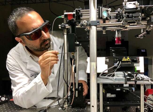
A paper on the work, All-optical Reflection-mode Microscopic Histology of Unstained Human Tissues, was published Sep.t 16 in the journal Scientific Reports.
Doctors now rely primarily on pre-operation MRI images and CT scans, experience and visual inspection to determine the margins of tumors during operations.
Tissue samples are then sent to labs for testing, with waits of up to two weeks for results to show if the tumor was completely removed or not.
In about 10 per cent of cases – the rates for different kinds of cancer involving tumors vary widely – some cancerous tissue has been missed and a second operation is required to remove it.
The photoacoustic technology developed at Waterloo works by sending laser light pulses into targeted tissue, which absorbs them, heats up, expands and produces soundwaves.
A second laser reads those soundwaves, which are then processed to determine if the tissue is cancerous or non-cancerous.
The system has already been used to make accurate images of even relatively thick, untreated human tissue samples for the first time ever, a key breakthrough in the development process.
Next steps include imaging fresh tissue samples taken during surgeries, integrating the technology into a surgical microscope and, finally, using the system directly on patients during operations.
“This will have a tremendous impact on the economics of health-care, be amazing for patients and give clinicians a great new tool,” said Haji Reza, director of the PhotoMedicine Labs at Waterloo. “It will save a great deal of time and money and anxiety.”
Researchers hope to develop a fully functioning system within about two years, a process including the need to clear ethical hurdles and securing regulatory approvals.
New intraoperative imaging techniques are being developed to provide an alternative to intraoperative histopathology methods.
Previous studies have used light sheet microscopy6, fluorescence nonlinear microscopy7,8,9,10, microscopy with ultraviolet surface excitation (MUSE) microscopy11, and structured light microscopy12,13 to recreate hematoxylin and eosin (H&E) histology images.
However, these techniques may require the addition of fluorescent dyes or optical clearing of the sample for imaging.
These labelling methods add logistic issues during an operation and introduce potential toxicities depending on the agent14,15.
Label-free approaches have been presented with stimulated Raman scattering microscopy16.
However, this technique has only been demonstrated with transmission-mode imaging which also requires thin samples and a complex and expensive picosecond scale (2 ps) dual pulse excitation.
Photoacoustic (PA) techniques take advantage of the large endogenous optical absorption contrast present in tissues.
As a nanosecond timescale pulse is absorbed by a chromophore it causes a sudden change in temperature.
This sudden change in temperature leads to transient thermoelastic expansion which in-turn generates acoustic waves in the ultrasound range.
Furthermore, biological tissue exhibits excellent specificity in optical absorption curves enabling rich tissue differentiation17,18,19.
For visualization of cell morphology, the ultraviolet (UV) absorption peak of DNA is a powerful target.
Conventional ultraviolet photoacoustic microscopy (UV-PAM) techniques have demonstrated efficacious label-free visualization of cellular structure in tissue samples20,21,22.
Indeed, some recent works have provided compelling histological imaging quality.
However, UV-PAM methods require physical contact with the tissue using an acoustic coupling medium, which may be impractical due to the constrained operational working space, increased risk of infection, and requires expensive pre- and post-surgical sterilization.
Also, in some UV-PAM devices the sample and portions of the apparatus were fully submerged in a water tank23.
There remains an unmet need for label-free cellular-scale imaging capable of providing real-time cellular structure visualization of optically thick (much thicker than transport mean free path) samples.
Thick samples require a reflection-mode apparatus which is more appropriate for clinical in-situ applications.
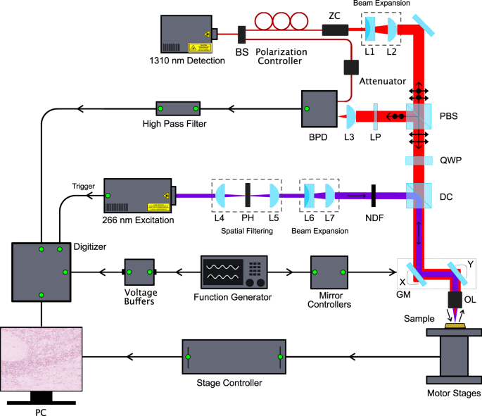
In this paper, we present an all-optical, fast, deep, label-free, non-contact, cellular-resolution, reflection-based optical imaging technique using the recently reported photoacoustic remote sensing (PARS) microscopy24,25,26.
A salient feature which sets PARS apart from conventional PA modalities is that it operates fully non-contact, even demonstrating centimeter scale working distances.
This permits optical absorption contrast to be visualised in turbid media without the need for acoustic coupling such as water or ultrasound gel.
The goal with this technique is to extract diagnostic quality histopathological information.
In PARS a nanosecond excitation beam is co-focused with a continuous-wave probe beam into the target.
The absorbed optical energy from the excitation pulse is converted to pressure through thermo-elastic expansion.
This pressure rise produces elasto-optic modulations within the absorber, changing the intensity of the back-reflected probe beam.
The magnitude of these optical signals is proportional to the optical absorption of the excitation wavelength27,28.
Utilizing the UV absorption peak of DNA, the cell nuclei and bulk tissue structure can be visualized. Employing PARS detection, we present the first label-free non-contact histology-like images of breast, gastrointestinal, and skin tissues from formalin-fixed paraffin embedded (FFPE) blocks and unstained thin slices.
These images are qualitatively compared by imaging co-localized regions and quantitatively compared by several nuclear morphology metrics to conventional H&E prepared slides to evaluate the diagnostic potential of this approach.
More information: Saad Abbasi et al. All-optical Reflection-mode Microscopic Histology of Unstained Human Tissues, Scientific Reports (2019). DOI: 10.1038/s41598-019-49849-9
Journal information: Scientific Reports
Provided by University of Waterloo


