


 Extract (6-MSITC) in Healthy Older Adults")
: An In-Depth Exploration into its Thermogenic Role and Social Significance")


One-third of the world population is infected with Toxoplasma, which causes a common eye infection called ocular toxoplasmosis.
Researchers have shed new light on how an infection with the parasite causes a distinctive lesion in the retina.
An international research team, from Flinders University in South Australia and University of São Paulo in Brazil, have identified proteins produced from infected retinal cells that push neighboring uninfected retinal cells to overgrow and create a distinctive lesion that doctors can use to diagnose the infection.
This study, published today in Microorganisms, is the first to use laboratory methods to understand how Toxoplasma infection leads to a characteristic eye lesion, after researchers monitored the process at a Brazilian eye clinic serving a region of 1.7 million people.
Flinders Strategic Professor in Eye & Vision Health and Superstar of STEM Justine Smith says the research demonstrates how retinal cells respond to an infection with Toxoplasma, and shows that the cell response may help parasites spread through the retina.
“In one of the largest groups of people with ocular toxoplasmosis studied to date, we see that infection causes a typical lesion in over 90 percent of infected eyes,” says Professor Smith.
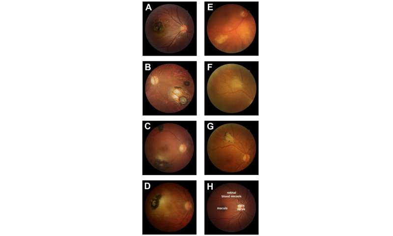
“Our findings show that T. gondii-infected human retinal pigment cells secrete VEGF and IGF1, and reduce production of TSP1, which promotes the proliferation of uninfected cells and renders those cells more susceptible to infection as a result.”
Dr. Joao Furtado, co-investigator from University of São Paulo and the International Agency for Prevention of Blindness, speculates that manipulating proteins within the eye might have therapeutic applications to limit the severity of the disease.
“There is no cure for toxoplasmosis, which is most often foodborne.
The World Health Organisation recommends food safety measures that include hand-washing, use of clean water in food preparation, and proper cooking,” advises Dr. Furtado.
Ocular toxoplasmosis is the most common cause of posterior uveitis in many countries, occurring subsequently after systemic infection with an obligate, intracellular, protozoan parasite, Toxoplasma gondii (T. gondii) [1].
Members of the feline family are the organism’s definitive hosts while humans and hundreds of other species may serve as intermediate hosts. Transmission to humans occurs by three principal routes: ingestion of raw or inadequately cooked infected meat, ingestion of oocysts (the environmentally resistant form of the organism defecated by cats that is found in cat litter and soil), and via vertical transmission [2].
T. gondii has a high affinity for the retinal microvascular endothelium with the retina being the primary site of infection in the eye [1,3].
Primary toxoplasmic retinochoroiditis can be defined as creamy-white exudative focal retinochoroiditis not associated with pre-existing retinochoroidal scars in either eye [4]. Fundus lesions of ocular toxoplasmosis at presentation can be characterized as primary or recurrent and active or inactive.
Chorioretinal lesions in Toxoplasma infection of the eye can occur either due to congenital or acquired infection [5].
Worldwide, the major victimized demographic in ocular toxoplasmosis continues to be congenitally infected fetuses and newborns, up to 95% of which may show retinochoroiditis that is mostly bilateral and recurrent [6,7]. Unilateral lesions are more common with acquired toxoplasmosis [8]. Lesions in immunocompetent individuals are less severe as compared to those in immunocompromised individuals [1].
Choroidal neovascular membrane (CNVM) is a late complication of ocular toxoplasmosis, mostly occurring in healed, inactive lesions and may be a cause of sudden loss of vision, especially in young patients [1,6,9]. Inflammatory CNVM in active toxoplasmic retinochoroiditis is a rare finding, having recently been reported by Hedge et al. However, factors in favour of an acquired or congenital aetiology of the ocular toxoplasmic lesions were not highlighted in the study [10].
We report the clinical presentation, diagnosis, and treatment of an immunocompetent adult with unilateral, primary, active toxoplasmic retinochoroiditis of an acquired aetiology that was complicated by early choroidal neovascularization. This case is from Pakistan, where no literature exists on ocular toxoplasmosis.
More information: Shervi Lie et al. Molecular Basis of The Retinal Pigment Epithelial Changes That Characterize The Ocular Lesion in Toxoplasmosis, Microorganisms (2019). DOI: 10.3390/microorganisms7100405
Provided by Flinders University


