


 Extract (6-MSITC) in Healthy Older Adults")
: An In-Depth Exploration into its Thermogenic Role and Social Significance")


Gone are the days when people use smart speakers – like Amazon Echo or Google Home – only as kitchen timers or dinner party music players.
These devices have started helping people track their own health, and can even monitor for cardiac arrest.
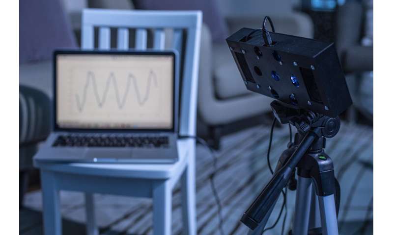
Now researchers at the University of Washington have developed a new smart speaker skill that lets a device use white noise to both soothe sleeping babies and monitor their breathing and movement.
With this skill, called BreathJunior, the smart speaker plays white noise and records how the noise is reflected back to detect breathing motions of infants’ tiny chests.
When the researchers tested BreathJunior with five babies in a local hospital’s neonatal intensive care unit, it detected respiratory rates that closely matched the rates detected by standard vital sign monitors.
The team will present its findings October 22 at the MobiCom 2019 conference in Los Cabos, Mexico.
“One of the biggest challenges new parents face is making sure their babies get enough sleep. They also want to monitor their children while they’re sleeping.
With this in mind, we sought to develop a system that combines soothing white noise with the ability to unobtrusively measure an infant’s motion and breathing,” said co-author Dr. Jacob Sunshine, an assistant professor of anesthesiology and pain medicine at the UW School of Medicine.
To make things easy for new parents, the team made a system that could run on a smart speaker that replicates the hardware in an Amazon Echo.
“Smart speakers are becoming more and more prevalent, and these devices already have the ability to play white noise,” said co-author Shyam Gollakota, an associate professor in the UW’s Paul G. Allen School of Computer Science & Engineering and the director of the UW computing for health group.
“If we could use this white noise feature as a contactless way to monitor infants’ hand and leg movements, breathing and crying, then the smart speaker becomes a device that can do it all, which is really exciting.”
White noise is a combination of different sound frequencies, which makes a seemingly random soothing sound that can help cover up other noises that might wake a sleeping baby.
To use white noise as a breathing monitor, the team needed to develop a method to detect tiny changes between the white noise a smart speaker plays and the white noise that gets reflected back from the infant’s body into the speaker’s array of microphones.
“We start out by transmitting a random white noise signal. But we are generating this random signal, so we know exactly what the randomness is,” said first author Anran Wang, a doctoral student in the Allen School.
“That signal goes out and reflects off the baby. Then the smart speaker’s microphones get a random signal back.
Because we know the original signal, we can cancel out any randomness from that and then we’re left with only information about the motion from the baby.”
Detecting breathing in babies has an extra wrinkle: the movement of their chests is so tiny that the smart speaker needs to know exactly where the babies are to be able to “see” them breathing.
University of Washington researchers have developed a new smart speaker skill that lets a device use white noise to both soothe sleeping babies and monitor their breathing and movement. Credit: University of Washington
“The breathing signal is so weak that we can’t just look for a change in the overall signal we get back,” Wang said.
“We needed a way to scan the room and pinpoint where the baby is to maximize changes in the white noise signal.
Our algorithm takes advantage of the fact that smart speakers have an array of microphones that can be used to focus in the direction of the infant’s chest.
It starts listening for changes in a bunch of potential directions, and then continues the search toward the direction that gives the clearest signal.”
BreathJunior tracks both small motions – such as the chest movement involved in breathing – and large motions – such as babies moving around in their cribs.
It can also pick up the sound of a baby crying.
The team created a prototype smart speaker to test BreathJunior on an infant simulator.
The researchers could set the simulator to breathe at specific rates, which allowed them to test how well BreathJunior detected a variety of respiratory rates – from a slow 20 breaths per minute to 60 breaths per minute.
The infant simulator also allowed the team to test if BreathJunior could detect abnormal breathing patterns, such as apnea, that are common in babies who are born early and may not have developed respiratory centers in their brains.
The system performed well for both tests.
Then the team tested how well their prototype tracked real babies’ breathing in the neonatal intensive care unit or NICU.
These babies are connected to wired, hospital-grade respiratory monitors, so the team could compare their readouts to BreathJunior’s.
The system was able to accurately identify respiratory rates up to 65 breaths per minute.
“Infants in the NICU are more likely to have either quite high or very slow breathing rates, which is why the NICU monitors their breathing so closely,” Sunshine said.
“BreathJunior holds potential for parents who want to use white noise to help their child sleep and who also want a way to monitor their child’s breathing and motion.
It also has appeal as a tool for monitoring breathing in the subset of infants in whom home respiratory monitoring is clinically indicated, as well as in hospital environments where doctors want to use unwired respiratory monitoring.
“However, it is very important to note that the American Academy of Pediatrics recommends not using a monitor that markets itself as reducing the risk of sudden infant death syndrome, and this research and the team makes no such claim.”
While BreathJunior currently uses white noise to track breathing and motion, the researchers would like to expand its capabilities so that it could also use other soothing sounds like lullabies.
The team plans to commercialize this technology through a UW spinout, Sound Life Sciences, Inc.
“In just a few years, we have come a long way from monitoring large motions in adults to extracting the tiny motion of a newborn infant’s breathing,” Gollakota said.
“This has been possible because of algorithmic innovations as well as advances in smart speaker hardware. Looking ahead, one can envision transforming a smart speaker into a ‘medical tricorder’ that can contactlessly monitor a variety of vital signs beyond just breathing.”
Apnea in infants is the term used to describe episodes of cessation of breathing and may be due to many physiological and pathophysiological processes. Brief periods of apnea that occur in short cycles of 5 seconds to 10 seconds is not pathologic and is referred to as periodic breathing. Periodic breathing is seen predominantly during the age of two to four weeks and resolves by age six months.[1][2][2]
Apnea is frequently seen in preterm infants but can occur at any age.
Apnea of prematurity is defined as a sudden cessation of breathing that lasts for at least 20 seconds or is accompanied by bradycardia or oxygen desaturation (cyanosis) in an infant younger than 37 weeks’ gestational age.
Apnea of infancy is defined as “an unexplained episode of cessation of breathing for 20 seconds or longer, or a shorter respiratory pause associated with bradycardia, cyanosis, pallor, and/or marked hypotonia.”
Apnea may be central, obstructive, or mixed.
Central apnea is due to the depressed respiratory center where there is a cessation of output from the central respiratory centers, and there is no respiratory effort.
Obstructive apnea occurs when there is an obstruction to the airway, and respiratory efforts are inadequate to maintain ventilation.
Mixed apnea (a period of central apnea, typically followed by airway obstruction) is the most frequent type among preterm infants.
Etiology
The etiology of apnea in infants is broad and varies according to the age of the infant and the pathophysiological mechanism.[3][4][5]
Preterm infants, especially those under 28 weeks gestation are highly prone for apnea due to the poor development of mechanisms of respiratory control and have apnea of prematurity.
Apnea soon after birth can occur due to birth asphyxia, maternal drug use, infections, metabolic causes and congenital anomalies.
Causes of Central apnea include central nervous system (CNS) infections (meningitis, encephalitis), head trauma (birth asphyxia or abusive trauma), toxin exposure, pertussis, infant botulism, inborn errors of metabolism (mitochondrial disease, Pompe disease, Leigh syndrome, and the mucopolysaccharidoses), metabolic derangements (hypoglycemia, hypocalcemia, and acidosis) and congenital anomalies (congenital central hypoventilation, Down syndrome and Arnold-Chiari malformation).
Obstructive apnea can occur due to obstructive sleep apnea, infections (pneumonia, croup), vocal cord paralysis, and congenital upper airway anomalies (e.g., Pierre-Robin sequence)
Mixed apnea occurs predominantly among premature infants but can also be caused by gastroesophageal reflux, pertussis, and bronchiolitis.
Epidemiology
The true prevalence and incidence of apnea in infants in unknown. Among preterm infants, the incidence of apnea is inversely related to gestational age with almost every infant younger than 28 weeks gestation having an episode and 50% of infants born between 33 weeks and 34 weeks. The incidence of apnea in the full-term infant is one per 1000.[6]
Pathophysiology
The relatively immature respiratory center among infants, particularly the preterm infants, makes them vulnerable to apneic episodes in the face of internal and external stressors. Unlike adults and older children, neonates respond to hypoxia and hypercarbia with a brief increase in respiratory rate followed by respiratory depression and apnea.
When feeding, poor coordination of sucking and breathing affects ventilation, further accentuated by exaggerated laryngeal chemical reflexes that depress the respiratory center causing apnea. Infants are very sensitive to stressors such as anemia, hypoglycemia, hypothermia, and toxin exposure that can depress the respiratory center.
The very pliable thoracic cage causes a collapse of the chest wall resulting in increased work of breathing and eventual tiring of chest muscles that lead to respiratory failure and apnea.
History and Physical
The initial history should be aimed to differentiate between a true apneic episode and periodic breathing or breath-holding spell. Once deemed as an apneic episode, a thorough review of the antenatal, perinatal, postnatal, and feeding history should be done. History of apneic episodes should be elicited as this indicates a life-threatening underlying cause and a higher probability of recurrence.
The family history of seizures, infant deaths, and the presence of serious illnesses in family members should also be ascertained. Social history should ask about potentially toxic exposures, including drugs or medications in the home, tobacco smoke exposure, and potential carbon monoxide exposure.
Associated symptoms such as sleep problems, snoring, mouth breathing should be inquired. A physical exam often provides valuable clues and aids to identify congenital anomalies, genetic syndromes or stigmata of inborn errors of metabolism and congenital infections.
Fever or hypothermia can raise the suspicion of sepsis or other infectious processes, tachypnea may indicate a lower respiratory tract infection or metabolic acidosis, and stridor indicates an upper airway obstruction. Unexplained skin bruises should raise a suspicion of child abuse.Go to:
Evaluation
The performance of lab and imaging studies should be guided by the history and physical exam findings. In neonates, complete blood count, serum glucose, calcium, and electrolyte measurement can be considered. If the infant is febrile or hypothermic and a serious infection is suspected, appropriate cultures of blood, urine, and possibly cerebrospinal fluid (CSF) should be obtained.
An EKG can be done to rule out cardiac dysrhythmias, especially the Long QT syndrome. Neuroimaging, EEG, specialist consults are not routinely recommended unless specifically indicated by the clinical picture. If the patient meets low-risk criteria for a Brief Resolved Unexplained Event (BRUE), no lab studies are indicated.[7][8]
Treatment / Management
The initial step in the management of apnea in infants is to assess the need for immediate resuscitation and/or stabilization of the infant. Subsequent management consists of determining the underlying etiology and instituting targeted specific therapy to the identified cause. For neonates, a period of observation with cardiorespiratory and pulse oximetry monitoring in the neonatal intensive care unit (NICU) is recommended. For infants with apnea of prematurity, interventions are recommended if apneic spells are frequent, prolonged, need frequent stimulation, or are associated with bradycardia and hypoxia. These infants benefit from nasal continuous positive airway pressure (CPAP) and methylxanthine therapy. There is no evidence that methylxanthine therapy is effective in term infants. Speech or occupational therapy may be consulted for infants with feeding-related issues. Management of infants in the low-risk BRUE category consists of parental reassurance and education. [9][10][11]
More information: Anran Wang et al, Contactless Infant Monitoring using White Noise, The 25th Annual International Conference on Mobile Computing and Networking – MobiCom ’19 (2019). DOI: 10.1145/3300061.3345453
Provided by University of Washington


