


 Extract (6-MSITC) in Healthy Older Adults")
: An In-Depth Exploration into its Thermogenic Role and Social Significance")


A study led by SMU suggests that oleandrin – a drug derived from the Nerium oleander plantcould prevent the HTLV-1 virus from spreading by targeting a stage of the reproduction process that is not currently targeted by existing drugs.
That is significant because there is currently no cure or treatment for the virus – a lesser-known “cousin” of HIV that affects an estimated 10 to 15 million people worldwide.
“Our research findings suggest that oleandrin could possibly limit the transmission and spread of HTLV-1 by targeting a unique stage in the retroviral life cycle,” said Robert Harrod, associate professor and director of Graduate Studies in SMU’s Department of Biological Sciences. Harrod is a co-author of the study, published in the Journal of Antivirals & Antiretrovirals.
The human T-cell leukemia virus type-1, or HTLV-1, is a retrovirus that infects white blood cells known as T-cells and is usually transmitted in a similar manner to HIV-1 through a person’s blood or body fluid.
Infected cells present within breast milk can also pass HTLV-1 from mother to infant through breastfeeding.
While HIV-1 kills the infected T-cells, HTLV-1 causes them to divide uncontrollably.
This in turn can lead to the development of aggressive leukemia – a cancer of the white blood cells. People infected with HTLV-1 can also develop a progressive neurological disease known as HTLV-1-associated myelopathy/tropical spastic paraparesis (HAM/TSP), a progressive inflammatory disease of the nervous system that can affect one’s ability to walk and may cause serious symptoms leading to coma and even death.
Retrovirus particles copy themselves within infected cells by transcribing their RNA into DNA after entering a cell, a process called the retroviral life cycle.
The more virus-infected cells that are produced, the worse symptoms can get for people who are infected with HTLV-1.
The two lead authors, Tetiana Bowley and Lacin Yapindi, are Ph.D graduate students who worked with Harrod in his lab. Aditi Malu, who also worked in Harrod’s lab, graduated from SMU with a Ph.D. in May.
Together with collaborator Jagan Sastry at the University of Texas M.D. Anderson Cancer Center and Dr. Robert Newman at Phoenix Biotechnology, Inc., SMU researchers found that the botanical compound called oleandrin successfully interrupted part of the infection cycle for HTLV-1.
“As has been shown for HIV-1, treatment with oleandrin did not affect the ability of infected cells to produce and release new virus particles. However, the particles that were produced were defective, meaning they contained less envelope glycoprotein on their surface,” Harrod said.
“This impaired their ability to form virological synapses for effective cell-to-cell virus transmission.”
A so-called “envelope,” which forms the outer coat of the HTLV-1 particle and binds to the receptors on the surface of target cells, must be present in order for a virus-infected cell to fuse with the membrane of an uninfected T-cell, allowing the virus to enter the cell and spread the disease. Without it, the HTLV-1 retrovirus can’t successfully be passed to other cells.
“Oleandrin is unique in its ability to block the incorporation of the envelope glycoprotein into mature virus particles as they’re exiting an infected cell,” Harrod said.
The hope is that oleandrin, or a similar drug that targets the same part of the retrovirus infection cycle, could potentially prevent HTLV-1 from causing progressively worse clinical symptoms in people with an immune-driven condition like HAM/TSP where the body’s immune system causes tissue damage due to the misrecognition of replicating virus particles.
“If a drug, such as oleandrin, could prevent the spread of HTLV-1 particles within an infected HAM/TSP patient, it may become possible to dampen the neuroinflammatory response to alleviate the symptoms of disease,” Harrod said.
Harrod called the findings “exciting” because oleandrin targets a different mechanism of fighting the virus—one that hasn’t been the focus of other antiviral drugs that attack specific steps in the retroviral infection cycle.
Those drugs, called highly-active antiretroviral therapies or HAART for short, have not been shown to be effective with HTLV-1.
In the study, to demonstrate that purified oleandrin or an N. oleander extract could inhibit the formation of HTLV-1 virological synapses, SMU researchers in Harrod’s lab labeled an HTLV-1-infected virus-producing cell-line with green fluorescent protein (GFP), so these cells could be easily identified by their ‘green’ fluorescence under a microscope.
These cells were then placed in the same culture well as healthy T-cells. T-cells that became infected with HTLV-1 were easy to spot because researchers could see a junction between the two cells and then a red fluorescent signal showing up in the newly-infected T-cell.
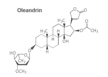
Nerium oleander (N. oleander) is an evergreen shrub that is frequently grown as an ornamental plant in gardens and public areas. N. oleander has linear and leathery leaves that come in various colours, from dark green to grey green with distinct light yellowish veins. Its flowers are fragrant, funnel-shaped and arranged in clusters at the tip of twigs, with white to pink to deep red colours. The fruit is a narrow pod containing many silky-haired seeds (Fig. 1). This plant is native to Mediterranean regions of Africa and Europe (1).

Nerium oleander (common oleander).
N. oleander is well known for its toxicity, as all parts of the plant contain numerous toxic compounds. The major toxic components are the cardiac glycosides oleandrin and neriin. Plants with red flowers produce more cardiac glycosides than plants with white flowers, especially in the flowering stage (2). Commonly, animal poisoning occurs due to accidental contamination of food or, in some cases, due to consumption of toxic plants by hungry animals (3). Cardiac glycosides inactivate the Na+/K+ ATPase pump on the cytoplasmic membrane of cardiac cells (4).
Despite its toxicity, N. oleander has been used as an abortifacient and for the treatment of heart failure, leprosy, malaria, ringworm and indigestion (5). N. oleander has insecticidal, molluscicidal, rodenticidal and antibacterial effects (6).
The lethal doses of N. oleander leaves were found to differ among animal species, such as sheep and rats (250 and 4,000 mg/kg body weight, respectively) (7,8). It has been reported that consuming minute amounts of oleander leaves, even 0.005% of the animal’s body weight, is fatal in cattle and horses (3).
The aim of this study was to evaluate the toxic effects of an ethanolic N. oleander leaf extract on haematological, cardiac, inflammatory, and serum biochemical parameters, as well as histopathological changes in the heart, after oral administration for 14 and 30 days.
MATERIALS AND METHODS
Plant material and preparation of the ethanolic extract
Fresh mature green leaves from N. oleander bushes with pink flowers were collected from 5 different locations on the Suez Canal University (Ismailia, Egypt) campus during February 2015. The leaves were washed with distilled water, dried in a hot-air oven at 40°C and then ground to minute pieces. Exactly 500 g of ground leaves was extracted several times with 90% ethanol using a separating funnel. The extract was filtered through filter paper (No. 1) and then evaporated under reduced pressure at 42°C using a rotary evaporator (RE111, BUCHI Corporation, DE, USA). The extract was stored in a 5°C refrigerator for further phytochemical analysis and toxicological study (9).
Phytochemical analysis
Moisture, ash and crude fibre content of the dried leaves were determined. Moisture and ash contents were estimated by subjecting 100 g of dried leaves to 90°C and 450°C for 12 hr and for 5 min, respectively. The percentage of crude fibre was measured according to the standards of the Association of Official Analytical Chemists (10).
In addition, glycosides and oleandrin (the most important bioactive constituents) in the extract were determined and calculated as percentages in 100 g of dried leaves. The total glycoside content was evaluated using Baljet’s reagent according to the method described by El-Olemy et al (11). After purification of the extract by lead acetate, Na2HPO4 and filtration, Baljet’s reagent was added, and the intensity of the produced reddish colour was then measured at 495 nm using a Shimadzu UV-2600 spectrophotometer. The concentration was calculated using a digitoxin standard curve (0.1–1.6 mg/mL). Oleandrin was extracted from N. oleander ethanolic extract with 1-naphthoyl chloride and quantified using HPLC (Agilent Infinity 1260) with a fluorescence detector (excitation = 220 nm and emission = 345 nm) equipped with a C18 reverse column according to the method described by Tor et al (12). The oleandrin standard was purchased from Sigma Chemical Co (St. Louis, MO, USA).
Acute oral toxicity (LD50 evaluation)
The median lethal dose (LD50) of the N. oleander ethanolic extract was determined using 12 BALB/c male mice. The animal management protocol and that experimental design were accepted by the Research Ethics Committee of the Faculty of Veterinary Medicine, Suez Canal University (Approval number 2016090). Graduated doses between 0.5 and 3.0 g of dried extract/kg b.w. were used after reconstitution in 0.5 mL of saline and administered orally by stomach gavage. The animals were observed for 24 hr for apparent signs of toxicity and mortality. The LD50 for the extract was determined according to the modified up-and-down technique (13), and its value was calculated using the acute oral toxicity statistical program (AOT425, version 1.0, USEPA, Washington DC, USA).
Animals and experimental design
A total of 30 male mice with body weights of 20–25 g were purchased from the Laboratory Animal Centre of the Faculty of Veterinary Medicine, Suez Canal University. The mice were housed in polypropylene cages (10 mice/cage) in a ventilated room with a controlled light (12 hr): dark (12 hr) cycle and temperature (25 ± 2°C). Food and water were provided ad libitum. The animal management protocol and the experimental design were accepted by the Research Ethics Committee of the Faculty of Veterinary Medicine, Suez Canal University (Approval number 2016090).
After one week of acclimatization, mice were randomly divided into 3 different groups: 10 animals in each group. Of these 10 mice, 5 were used for the 14-day study, and the other 5 were used for the 30-day experiment. The mice in the 1st group were administered normal saline and kept as controls. The animals in the 2nd and 3rd groups were administered N. oleander ethanolic extract using intragastric gavage at doses of 100 and 200 mg dried extract/kg body weight, respectively, in 0.5 mL of saline. These doses were determined to be less than one tenth (1/10) of the calculated LD50 in this study (14) to avoid any possible toxin accumulation in the mice. Nerium extract was administered daily for 14 and 30 consecutive days from the beginning of the treatment.
Blood collection, serum and tissue preparation
After 14 and 30 days of oral administration of N. oleander, blood samples were collected from the postorbital plexus 24 hr after the last treatment. The blood samples were collected in EDTA tubes to investigate the haematological parameters and in non-EDTA-containing tubes to prepare serum for biochemical analysis.
The mice were sacrificed by decapitation, and heart samples were rapidly excised for histopathological evaluation.
Blood and serum biochemical analysis
Blood parameters, including red blood cells (RBCs), haemoglobin concentration (Hb), haematocrit value (HCT), mean corpuscular volume (MCV), mean corpuscular haemoglobin (MCH), mean corpuscular haemoglobin concentration (MCHC), white blood cells (WBCs), lymphocytes, and platelet count (PLT), were estimated. Alanine aminotransferase (ALT), creatinine, and inflammatory markers, including interleukin 1 (IL-1), interleukin 6 (IL-6), tumour necrosis factor α (TNF-α) and C reactive protein (CRP), were measured in serum. Myocardial damage markers, including lactate dehydrogenase (LDH), creatine kinase (CK) and creatine kinase MB (CK-MB), were also evaluated. ALT, creatinine, CRP, CK, CK-MB and LDH were measured using a Roche/Hitachi Cobas C311 automatic analyzer. Each parameter was analysed according to the method and the instructions provided by the manufacturer of the automated analyzer. The CBC was analysed using an automated Sysmex KX-21N haematology analyzer according to the manufacturer’s instructions. TNF, IL-1 and IL-6 were estimated by an ELISA reader in accordance with the manufacturer’s directions. All machines and equipment were calibrated and standardized periodically before running the tests.
Histopathological evaluation
Heart muscles were washed with normal saline solution (0.9% NaCl in distilled water) and then fixed in 10% buffered formalin. After standard processing of the tissue, 5 μm sections were prepared and stained with haematoxylin and eosin. The slides were then examined under a light microscope to investigate the histopathological changes. All images were captured with a calibrated standard digital microscope camera (ISH1000 digital microscope camera, Fuzhou Tucsen Photonics Co., Fujian, China) using an CX21 microscope (Olympus Co., Tokyo, Japan), with “IS Capture” software (Olympus Co.) for image capture and enhancement. A semi-quantitative scoring system for myocardial damage was applied based on the severity and extent of the lesions observed in each mouse (15). Briefly, for each myocardial slide, histopathological signs of inflammation and/or myocarditis were scored on a five-degree scale ranging from 0 to 4+. A score of zero indicated no or doubtful presence of lesions in each category. While a score of 1+ described a limited focal distribution of lesions, scores of 2+ to 3+ denoted intermediate rigorousness with multiple lesions, and a score of 4+ indicated the presence of coalescing and extensive lesions throughout the entire examined myocardial tissue. A maximum score of 16 was possible using this scoring system.
Statistical analysis
Data were examined using one-way ANOVA followed by Duncan’s post hoc test to detect significant differences at a probability level of 5% (p ≤ 0.05). All data were expressed as the mean ± SE.
RESULTS
Phytochemical analysis
Moisture, ash, crude fibre, total glycoside and oleandrin contents were estimated in dried leaves of N. oleander and are presented in Table 1. The total glycoside and oleandrin contents were 1,944 and 82 mg/100 gm dried leaves, respectively.
Table 1
Phytochemical analysis of Nerium oleander dried leaves
| Consistent of dried leaves | Percentage |
|---|---|
| Moisture | 5.13 |
| Ash | 9.06 |
| Crude fiber | 20.66 |
| Glycosides | 1.94 |
| Oleandrin | 0.0082 |
LD50 experiment results
Oral administration of N. oleander ethanolic extract up to a dose of 3 g/kg b.w. revealed neither toxic signs nor death within 24 hr of extract administration. Furthermore, there were no apparent signs of delayed toxicity when the mice were observed for an additional 14 days.
Blood parameter estimation results
After 14 days of oral treatment with N. oleander alcoholic extract, blood parameters showed a significant increase in MCH and a decrease in WBCs in the high-dose group, with a significant decrease in lymphocytes (%) in both the low- and high-dose groups. After 30 days of oral administration, blood parameters revealed significant elevations of MCV, WBCs and PLT in the high-dose group, with significant decreases in lymphocytes (%) in the low- and high-dose groups compared to those in the control group (Table 2, ,33).
Table 2
Blood parameters at 14 days post oral treatment with Nerium oleander ethanolic extract
| Parameter/Group | Control | Low dose | High dose |
|---|---|---|---|
| RBCs (106)/μL | 7.90 ± 0.20a | 7.52 ± 0.24a | 7.53 ± 0.30a |
| Hb g/dL | 10.78 ± 0.31a | 10.64 ± 0.21a | 10.52 ± 0.22a |
| HCT% | 38.50 ± 0.77a | 37.44 ± 0.46a | 36.80 ± 0.95 a |
| MCV fL | 48.38 ± 0.27a | 47.86 ± 0.48a | 47.98 ± 0.75a |
| MCH pg | 12.22 ± 0.23b | 12.76 ± 0.56b | 13.90 ± 0.55a |
| MCHC g/dL | 27.12 ± 0.45b | 27.72 ± 0.46ab | 28.86 ± 0.63a |
| WBC (103)/μL | 10.36 ± 0.34a | 10.12 ± 0.23a | 9.12 ± 0.49b |
| Lymphocyte % | 95.60 ± 0.97a | 93.20 ± 1.11b | 91.00 ± 0.91b |
| PLT (103)/μL | 955.60 ± 15.95a | 1031.00 ± 70.03a | 1059.20 ± 89.06a |
Data are expressed as means ± SEM, n = 5. Values having different alphabetic superscripts within the same row are significantly different (p ≤ 0.05).
Table 3
Blood parameters on 30 days post oral treatment with Nerium oleander ethanolic extract
| Parameter/Group | Control | Low dose | High dose |
|---|---|---|---|
| RBCs (106)/μL | 8.13 ± 0.53a | 8.17 ± 0.25a | 7.86 ± 0.33a |
| Hb g/dL | 11.72 ± 0.58a | 11.68 ± 0.53a | 11.14 ± 0.79a |
| HCT% | 41.02 ± 2.07a | 40.68 ± 0.77a | 42.28 ± 0.90 a |
| MCV fL | 49.14 ± 0.69b | 48.78 ± 0.94b | 53.78 ± 0.39a |
| MCH pg | 14.02 ± 0.37a | 14.28 ± 0.29a | 14.90 ± 0.47a |
| MCHC g/dL | 28.30 ± 0.34a | 29.82 ± 0.56a | 28.80 ± 0.91a |
| WBC (103)/μL | 10.70 ± 0.88b | 9.04 ± 1.04b | 14.14 ± 1.09a |
| lymphocyte % | 91.00 ± 2.12b | 95.00 ± 1.08a | 94.60 ± 0.88a |
| PLT (103)/μL | 771.00 ± 88.69b | 789.60 ± 66.51b | 1082.40 ± 129.07a |
Data are expressed as means ± SEM, n = 5. Values having different alphabetic superscripts within the same row are significantly different (p ≤ 0.05).
Serum biochemical parameter estimation results
IL-1, IL-6, TNF-α, CK, and CK-MB were significantly increased in the high-dose group after 14 days of treatment, while CRP and LDH were significantly increased in both the low- and high-dose groups. After 30 days of administration, IL-6, TNF-α, CRP, ALT, LDH, CK and CK-MB levels were significantly increased in both the low- and high-dose groups, while IL-1 was only significantly increased in the high-dose group compared to those in the control group (Table 4, ,55).
Table 4
Serum biochemical parameters after 14 days from oral treatment with Nerium oleander alcoholic extract
| Parameter/Group | Control | Low dose | High dose |
|---|---|---|---|
| IL1 pg/mL | 2.72 ± 0.26b | 2.76 ± 0.139b | 5.12 ± 0.440a |
| IL6 pg/mL | 9.46 ± 0.30b | 9.34 ± 0.320b | 15.42 ± 0.886a |
| TNF α pg/mL | 8.70 ± 0.24b | 9.12 ± 0.214b | 12.36 ± 0.594 a |
| CRP | 3.74 ± 0.37c | 5.72 ± 0.599b | 8.24 ± 0.315a |
| ALT | 44.20 ± 5.56a | 33.40 ± 2.221b | 47.80 ± 3.795a |
| Creatinine | 0.48 ± 0.05a | 0.48 ± 0.048a | 0.50 ± 0.041a |
| LDH | 54.00 ± 3.24c | 63.60 ± 2.183b | 84.20 ± 4.990a |
| CK | 237.20 ± 11.60b | 259.20 ± 15.489b | 426.40 ± 47.646a |
| CK Mb | 16.40 ± 1.20b | 20.20 ± 0.753b | 116.20 ± 7.973a |
Data are expressed as means ± SEM, n = 5. Values having different alphabetic superscripts within the same row are significantly different (p ≤ 0.05).
Table 5
Serum biochemical parameters after 30 days from oral treatment with Nerium oleander ethanolic extract
| Parameter/Group | Control | Low dose | High dose |
|---|---|---|---|
| IL1 pg/mL | 2.92 ± 0.29b | 3.66 ± 0.211b | 8.08 ± 0.511a |
| IL6 pg/mL | 9.44 ± 0.30c | 12.40 ± 0.623b | 18.74 ± 0.631a |
| TNF α pg/mL | 8.64 ± 0.28c | 11.24 ± 0.446b | 16.66 ± 0.290a |
| CRP | 3.84 ± 0.43c | 6.44 ± 0.431b | 9.00 ± 0.922a |
| ALT | 47.20 ± 4.03c | 58.20 ± 2.955b | 72.00 ± 5.745a |
| Creatinine | 0.50 ± 0.06ab | 0.40 ± 0.058b | 0.58 ± 0.048a |
| LDH | 53.80 ± 3.66c | 95.20 ± 1.844b | 116.40 ± 5.141a |
| CK | 239.80 ± 14.60c | 357.80 ± 29.786b | 550.40 ± 75.563a |
| CK Mb | 16.80 ± 1.18c | 26.00 ± 1.472b | 42.00 ± 4.546a |
Data are expressed as means ± SEM, n = 5. Values having different alphabetic superscripts within the same row are significantly different (p ≤ 0.05).
Histopathological results
In the control group, heart muscle formed of myocytes arranged into fascicles with striations and peripheral oval nuclei separated by loose connective tissue and thin vessels (Fig. 2A, 2D).

Histopathological results on 14 (A, B, C) and 30 days (D, E, F) post oral treatment with Nerium oleander ethanolic extract. All slides were stained with Hematoxylin and Eosin dyes and captured with high power field of 400×. Arrows and arrowheads point out the pathological changes such as inter-fascicular edema (black arrow) and degenerated myocytes with vacuolation of myofibrils (arrow head).
After 14 days of oral administration of a low dose of N. oleander alcoholic extract, heart muscles showed diffuse mild inter-fascicular oedema with mildly congested vessels. In addition, many degenerated myocytes showed fragmentation of myofibrils (Fig. 2B). In contrast, in the high-dose group, the heart muscles showed moderate inter-fascicular oedema with dilated congested vessels and few degenerated myocytes with focal striation loss and focal vacuolar degeneration (Fig. 2C).
After 30 days of oral administration of a low dose of N. oleander extract, the heart muscles showed focal mild inter-fascicular oedema with mildly congested vessels and very few degenerated myocytes (Fig. 2E). In contrast, in the high-dose group, the heart muscles showed focal marked inter-fascicular oedema with dilated congested vessels (black arrow) and moderately degenerated myocytes with vacuolation of the muscle. Additionally, a few myofibrils showed striation loss (arrow head) (Fig. 2F). Table 6 shows semi-quantitative histopathological scoring of mouse cardiac lesions at 14 and 30 days after N. oleander extract administration.
Table 6
Semi-quantitative histopathological scoring of myocardial damage after 14 and 30 days from oral treatment with Nerium oleander alcoholic extract in mice
| Groups | 14 days | 30 days |
|---|---|---|
| Control | 0.0 ± 0.00b | 0.2 ± 0.26c |
| Low dose | 2.0 ± 0.41a | 2.6 ± 0.32b |
| High dose | 3.2 ± 0.75a | 4.6 ± 0.66a |
Data are expressed as means ± SEM, n = 5. Values having different alphabetic superscripts within the same column are significantly different (p ≤ 0.05).Go to:
DISCUSSION
Oleander poisoning is a common problem in many parts of the world.
Oleander toxicity is due to oleandrin (the main cardiac glycoside of oleander) and neriin, which cause damage by inhibiting the plasmalemmal Na+/K+-ATPase. Na+/K+-ATPase is responsible for maintaining the membrane potential of cardiac muscle cells, and its inhibition leads to necrosis of myocytes (16).
In the current study, we examined the haematological, biochemical and histopathological changes that occur after administration of an N. oleander leaf ethanolic extract in mice. Haematological analysis showed a significant increase in MCV, WBCs and PLT, and these results are consistent with the results of other researchers (17,18).
The increase in MCV is an indication of a pathological condition called “macrocytosis”, which is due to liver injury that may be caused by free radicals resulting from N. oleander administration (18).
The liberation of free radicals into the blood may also influence the circulating cells and significantly altera their numbers (19). Significant decreases in RBC, Hb and PCV were recorded in female mice treated with N. oleander alkaloid extracts (20). The author attributed these declines to the disruption of factors that influence erythropoiesis or to the rupture of the RBC membrane. However, the effect of N. oleander on blood parameters remains controversial among researchers (18,20). Higher platelet and white blood cell counts may act as markers of pro-inflammatory conditions (21).
These results revealed significant increases in IL-6, TNF-α, CRP, ALT, LDH, CK and CK-MB levels in both the low- and high-dose groups, while IL-1 was significantly increased only in the high-dose group compared to the control group.
Increased serum concentrations of the inflammatory marker CRP and pro-inflammatory cytokines (IL-1, IL-6 and TNF-α) are observed during chronic heart failure and mediate both immune and inflammatory responses, inducing apoptosis, in myocardial cells (22) (23). Toxic substances induce pro-inflammatory cytokines through the activation of macrophages (24).
The pro-inflammatory cytokine TNF-α is involved in the initiation and progression of the inflammatory response and the production of inflammatory mediators, such as IL-1 and IL-6 (25–27). Our findings are consistent with the results obtained by other investigators (28,29).
The liver enzyme (ALT) was elevated in the serum, especially after 30 days of N. oleander treatment, indicating liver injury and disruption of hepatocyte membranes, which may be due to the liberation of free radicals by N. oleander (18,30). LDH levels were also significantly increased, which could represent a good marker along with ALT, of tissue and cellular damage, especially hepatic cells.
CK is an essential enzyme in cellular energy transfer, helping in the transfer of high-energy phosphoryl groups in the mitochondria to generate ATP (31). The MB isoform of creatine kinase (CK-MB) is considered to be a specific indicator of myocyte injury and is elevated in myocardial damage (32). Jortani et al. (33) reported that oleander plants containing the cardiac glycoside oleandrin exert toxic effects via inhibition of Na+/K+-ATPase pump activity.
In this study, CK and CK-MB were significantly elevated, indicating severe myocardial damage that may be due to cardiac glycoside inhibition of the Na+/K+-ATPase pump in the cytoplasmic membrane of cardiac cells, leading to positive inotropic effects and spontaneous depolarization in cardiac muscles (34).
The inhibition of Na+/K+-ATPase leads to an increase in the intracellular sodium concentration, which in turn induces an increase in the intracellular calcium concentration via the sodium-calcium exchanger. The sodium-calcium exchanger is particularly active in the myocardium and in vascular smooth muscles. The increase in intracellular calcium increases the force of contraction of the heart and vascular smooth muscles (35).
Our results were confirmed by histopathological examinations, which indicated several degrees of myocardial vacuolar degeneration, the loss of myofibril striation and inter-fascicular oedema. These findings indicate some degree of myocyte damage and myocardial injury after treatment with N. oleander, and these results are consistent with those observed by several researchers (1,36,37).
Finally, we can conclude that exposure to N. oleander leaf extract adversely affects the heart and liver. The results showed significant adverse changes in haematological, biochemical and inflammatory parameters, as well as histopathological alterations. As this plant is used in the medical field, extra effort must be made to purify the effective chemical constituents to avoid toxicity.
More information: Tetiana Hutchison et al. The Botanical Glycoside Oleandrin Inhibits Human T-cell Leukemia Virus Type-1 Infectivity and Env-Dependent Virological Synapse Formation, Journal of Antivirals & Antiretrovirals (2019). DOI: 10.35248/1948-5964.19.11.184


