


 Extract (6-MSITC) in Healthy Older Adults")
: An In-Depth Exploration into its Thermogenic Role and Social Significance")


In decades of studying how neural circuits in the brain’s visual cortex adapt to experience, MIT Professor Mark Bear’s lab has followed the science wherever it has led, yielding the discovery of cellular mechanisms serving visual recognition memory, in which the brain learns what sights are familiar so it can focus on what’s new, and of a potential therapy for amblyopia, a disorder where children born with disrupted vision in one eye can lose visual acuity there permanently without intervention.
But this time his lab’s latest investigation has yielded surprising new layers of mystery.
Heading into the experiments described in a new paper in Cerebral Cortex, Bear and his team expected to confirm that key proteins called NMDA receptors act specifically in neurons in layer 4 of the visual cortex to make the circuit connection changes, or “plasticity,” necessary for both visual recognition memory and amblyopia. Instead, the study has produced unexpectedly divergent results.
“There are two stories here,” said Bear, co-senior author and Picower Professor of Neuroscience in The Picower Institute for Learning and Memory. “One is that we have further pinpointed where to look for the root causes of amblyopia.
The other is that we have now completely blown up what we thought was happening in stimulus-selective response potentiation, or SRP, the synaptic change believed to give rise to visual recognition memory.”
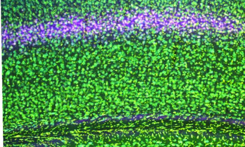
The cortex is built like a stack of pancakes, with distinct layers of cells serving different functions.
Layer 4 is considered to be the primary “input layer” that receives relatively unprocessed information that arises from each eye.
Plasticity that is restricted to one eye has been assumed to occur at this early stage of cortical processing, before the information from the two eyes becomes mixed.
However, while the evidence demonstrates that NMDA receptors in layer 4 neurons are indeed necessary for the degradation of vision in a deprived eye, they apparently play no role in how neural connections, or synapses, serving the uncompromised eye strengthen to compensate, and similarly don’t matter for the development of SRP.
That’s even though NMDA receptors in visual cortex neurons have directly been shown to matter in these phenomena before, and layer 4 neurons are known to participate in these circuits via telltale changes in electrical activity.
“These findings reveal two key things,” said Samuel Cooke, co-senior author and a former member of the Bear Lab who now has his own at King’s College London.
“First, that the neocortical circuits modified to enhance cortical responses to sensory inputs during deprivation or to stimuli that have become familiar reside elsewhere in neocortex, revealing a complexity that we had not previously appreciated.
Second, the results show that effects can be clearly manifest in a region of the brain that are actually echoes of plasticity occurring elsewhere, thereby illustrating the importance of not only observing biological phenomena but also understanding their origins by locally disrupting known underlying mechanisms.”
‘We were stunned’
To perform the study, Bear Lab postdoc and lead author Ming-fai Fong and used a genetic technique to specifically knock out NMDA receptors in excitatory neurons in layer 4 of the visual cortex of mice.
Armed with that tool, she could then investigate the consequences for visual recognition memory and “monocular deprivation,” a lab model for amblyopia in which one eye is temporarily closed early in life. The hypothesis was that knocking out the NMDA receptor in these cells in layer 4 would prevent SRP from taking hold amid repeated presentations of the same stimulus, and would prevent the degradation of vision in a deprived eye as well as the commensurate strengthening of the unaffected eye.
“We were gratified to note that the amblyopia-like effect of losing cortical vision as a result of closing an eye for several days in early life was completely prevented,” Cooke said. “However, we were stunned to find that the two enhancing forms of plasticity remained completely intact.”
Fong said that with continued work, the lab hopes to pinpoint where in the circuit NMDA receptors are triggering SRP and the compensatory increase in strength in a non-deprived eye after monocular deprivation. Doing so, she said, could have clinical implications.
“Our study identified a crucial component in the visual cortical circuit that mediates the plasticity underlying amblyopia,” she said.
“This study also highlights the ongoing need to identify the distinct components in the visual cortical circuit that mediate visual enhancement, which could be important both in developing treatments for visual disability as well as developing biomarkers for neurodevelopmental disorders. These efforts are ongoing in the lab.”
The search now moves to other layers, Bear said, including layer 6, which also receives unprocessed input from each eye.
“Clearly this is not the end of the story,” Bear said.
Amblyopia is a cortical developmental disorder, secondary to abnormal visual inputs to each eye occurring early in life (during the cortical plasticity stage) where in dissimilar action potentials (in amplitude or time, or both) generated in the retina reach the cortex.
These cortical changes entice the visual cortex to prefer one eye over the other, leading to a number of functional deficiencies in the eye, altered visual function like decreased vernier acuity, and impaired contrast sensitivity, particularly to detect high spatial frequency stimuli and impaired motor signs like hand-eye coordination and spatial localization, and it can be either unilateral or bilateral.[1]
Amblyopia is the most common cause of monocular visual impairment affecting 2–5% of the general population.
Prevalence estimates from population-based studies in children age 6–71 months range from 0.73 to 1.9%,[2] whereas school-based studies of older children typically report higher rates (range: 1.0–5.5%)[2] depending on the population studied and the definition used.[2,3,4,5,6,7,8,9,10,11,12,13,14]
Bilateral amblyopia is less frequent than unilateral, but the reported proportion varies considerably, from as low as 5% up to 41% of all cases of amblyopia,[4,11,12,13,14,15,16] and 2.7-18 times greater when strabismus is present.[12,15,17,18,19]
Risk factors for amblyopia are premature birth, small for gestational age,[20,21,22,23,24] developmental delay,[24] or having a first-degree relative with amblyopia.[25,26] Environmental factors, including maternal substance abuse during pregnancy, have been reported to be associated with an increased risk of amblyopia or strabismus in some studies.[27,28,29,30,31,32] However, some studies have refuted the same.[12,15,33]
In this review, we attempt to give an insight in to the various aspects of the normal cortical development during early postnatal life. The topic is covered in different headings to make it convenient for the practitioner, to comprehend the various treatment modalities, when to initiate, or terminate, and what to expect.Go to:
Anatomy and physiology of the retino-geniculo-cortical pathway
A clear understanding of the retino–geniculo–cortical path way is very essential for the proper understanding of amblyopia. We have included a brief note about the passage of electrical impulse from retinal ganglion cell to the primary visual cortex and beyond. In amblyopia, the cortical changes are not limited to V1 and extend beyond. The so-called critical period extends from birth to 7–8 years. Strabismus, refractive error, cataract, and ptosis, occurring during this critical period are highly amblyopiogenic.
The normal visual experience during the critical period is essential for the cortical development, and this continues postnatally till the age of 7 years. After this, the cortical plasticity decreases, but is never fully lost till early 50s.[34] The ganglion cell is the first step where the light energy is converted in to neural impulse.[35] Hence, for the purposes of this review, we shall confine ourselves from ganglion cells onwards.
Simply put, ganglion cells are of two types: parvocellular (P cells) and magnocellular (m cells) types.[35] The former are in the foveal and parafoveal areas and the latter in the perifoveal and peripheral areas of retina.
There are many direction-sensitive retinal ganglion cells, which fire when the image moves in one direction and not in the other direction.[35] P cells are involved in fine visual acuity, fine stereopsis, and color vision and M cells are involved in gross stereopsis and movement recognition.[35] P cells have a larger representation in the sensory cortical areas, compared with M cells.[36] There is again dichotomy of the ganglion cells.
Those axons of the ganglion cells on the nasal side of a line splitting the fovea (in to nasal and temporal halves) cross over to the opposite side and those on temporal side go to the geniculate nucleus on the same side.[36]
Retinal fibers end in the lateral geniculate nucleus (a part of the thalamus and distinctly stratified) in specific layers. The right and left fibers are distinctly separate. The parvocellular and magnocellular fibers end in different layers in the lateral geniculate nucleus. Nearly 90% geniculate afferents are concerned with the vision.[35,36]
For the purposes of discussion here, we shall ignore the rest of the 10% of the fibers. In the lateral geniculate nucleus, there is some modification of the stimulus from each eye, although topographical representation is exactly the same as retina. The adjacent retinal ganglion cells discharge on to the adjacent geniculate cells.Go to:
Cortical development
In their classical studies of normal visual development and the effects of deprivation, Hubel and Wiesel showed that the axons of relay cells in the lateral geniculate nucleus (LGN) that terminate in layer IV of the primate primary visual cortex (area 17) segregated over the first 3 weeks of life to form the ocular dominance columns (ODC) in which the inputs from the two eyes alternate equally.[37] However, if one eye was closed at birth, its ocular dominance bands became very narrow and those from the fellow eye expand.
This enlargement of the LGN cell bodies is thought to occur because they must sustain more extensive axonal arborizations in their enlarged ocular dominance bands in the visual cortex.
When closure of an eye was delayed until 6–7 months of age, this produced only a very small difference in size between cells in the deprived and undeprived LGN laminae after 2 months of closure indicating that there is a second sensitive period in which the pattern of changes produced by visual deprivation is different.[38]
The number of synapse in the primate visual cortex continues to increase until at least 6 months of age before progressively falling back to adult levels.[39] It may be the elaboration of these connections during development that leads to their increasing susceptibility to visual deprivation and the different effects of monocular deprivation at later ages.[40] Such connections are more important for parvocellular than magnocellular cells. In all likelihood, the shrinkage of parvocellular cell bodies in the LGN occurs because monocular closure prevents binocular cooperation and the cortical axonal arborizations of parvocellular cells related to both eyes make fewer connections and are smaller than normal.[41]
During the postnatal period, the cortical circuitry is not mature. The ocular dominance columns are amenable to alteration. Plasticity is a developmental necessity and the maturity of the ODC continues till the age of 36 months postnatally and during this maturation process visual experience from the two eyes must be matched[42]
The maturation is completed by the GABAergic interneurons in the layer 2 and 3 of V1.[43] These interneurons are inhibitory in nature and inhibition of these interneurons can theoretically prolong the plasticity period.[44] The maturation process makes the cortical circuitry immune to altered visual experience as happens after the age of 10 years. However, the cortical plasticity is never fully lost in adults, and if there are any ways of restoring plasticity of the cortical circuitry, amblyopia is curable.Go to:
Cortical changes causing amblyopia
Orssaud et al. in their article on amblyopia state that amblyopia is a developmental disorder of the entire visual system, including the extrastriate cortex, although it manifests as impaired visual acuity in the amblyopic eye, other abnormalities of visual function such as decreased contrast sensitivity and stereoscopic vision are observed, and some abnormalities can be found in the “good” eye. Since amblyopia occurs during the critical period of brain development, it may be due to organic pathology of the visual pathways, visual deprivation, or functional abnormalities, mainly anisometropia or strabismus.[45]
The suppression of the visual input from the weaker eye has been suggested as the underlying reason of the amblyopic syndrome, although it is still an unresolved question as to what extent neural responses to the visual information coming from the amblyopic eye are suppressed during binocular viewing.[46]
To address this question, Körtvélyes et al. measured event-related potentials (ERPs) to foveal face stimuli in amblyopic patients, both in monocular and binocular viewing conditions. They found early ERP components were reduced and delayed in the case of monocular stimulation of the amblyopic eye as compared with the fellow eye stimulation or to binocular viewing and the input from the amblyopic eye is completely suppressed already at the earliest stages of visual cortical processing during binocular viewing.Go to:
What are other features affected in amblyopic eye?
Prior studies by McKee et al. have found a reduction in contrast sensitivity in eyes with amblyopia using sinusoidal gratings, whereas minimal loss has been reported with Pelli-Robson charts. Most studies have evaluated contrast sensitivity at the time of diagnosis of amblyopia or after short-term treatment.[47] Repka et al. have evaluated the contrast sensitivity using Pelli-Robson low contrast letter charts at age 10 years, several years after treatment of amblyopia and found that the distribution of contrast sensitivity in the amblyopic eye was similar to that reported for monocular testing of normal 10-year olds.[48]
The younger the patients at enrollment into the randomized trial (3 to <5 years compared with those 5 to <7 years), the more likely to have slightly better contrast sensitivity in the amblyopic eye at 10 years of age This effect, if substantiated, could be due to a number of factors like a younger age at treatment allowing more complete cortical development, and a shorter duration of the vision deficit.
Each of these circumstances might allow a more complete treatment effect or alternatively a shorter period of insult and thus less profound insult to the developing visual sensory system. Nevertheless, it seems likely that mild residual amblyopia is associated with only a mild reduction in contrast sensitivity after treatment of moderate amblyopia from strabismus, anisometropia, or both.
Slyshalova in their study recorded different types of electroretinograms (ERG) as part of the International Society for Clinical Electrophysiology of Vision (ISCEV) standard, as well as macular (15%) pattern and multifocal ERG in 41 children aged 5–17 years, who had high amblyopia with varying gaze fixation and a visual acuity of 0.03–0.1. In high amblyopia, the mixed, macular, and flicker (30 Hz) ERGs were unchanged; however, some patients had supernormal a-wave of a mixed ERG, subnormal a-a- and b-waves of a macular ERG, and a moderately subnormal ERG pattern.
Recording of a multifocal ERG showed lower retinal density values in the first and second rings.[49] Karlica et al. correlated visual evoked potential (VEP) parameters (amplitude and latency) with visual acuity of the amblyopic eye and found that VEP may be a valid method to determine amblyopia.[50] Thus, in high amblyopia, there are characteristic retinal bioelectrical activity impairments recorded under different conditions of stimulation and adaptation, which suggest that there are impaired interreceptor relations at the retinal level. These changes statistically significantly differ from those in organic retinal defects, which may be a criterion for their differential diagnosis.
Effect on stereopsis: In individuals with amblyopia, the relationship between the visual acuity of the amblyopic eye and stereoacuity is complex, as illustrated by Fig. 1, replotted from a large-scale study.[51] Overall, worse visual acuity seems to correlate with worse stereoacuity. However, this relationship seems mostly driven by anisometropic subjects (blue symbols). Indeed, over the entire range of amblyopic eye visual acuities, there are amblyopes who are essentially stereo-blind (red and gray symbols plotted along the top of the graph).
These are mainly strabismic amblyopes, whether purely strabismic or mixed (strabismic and anisometropic). It is worth noting that constant strabismics with good acuity in both eyes are generally stereoblind.[52] However, recent work suggests that coarse stereopsis may be selectively spared in stereo-deficient children with a history of amblyopia.[53]
Fig. 1 indicates stereoacuity vs. visual acuity. The dotted lines show the upper and lower limits of the test. The data for strabismic anisometropes (gray squares) have been slightly displaced for clarity.[51] The blue regression line suggests that worse visual acuity goes hand in hand with worse stereoacuity in anisometropic amblyopes; however, this relationship does not hold in strabismic amblyopes or strabismic anisometropes.[51]
Are all amblyopias the same?
Classification of amblyopia is based on etiology, which is heterogeneous, may be caused by stimulus deprivation, strabismus, refractive error, or a combination of these. Amblyopia is usually unilateral, but it may be bilateral in cases of bilateral high refractive error or bilateral ocular pathology, such as cataract.[54]
Keech et al. in their paper mention that the commonest risk factors for amblyopia are constant strabismus and different refractive errors in each eye and age of the child when exposed to an amblyopia-inducing condition is the most important determinant for the development of amblyopia.[54]
Vojnosanit Pregl have published in their work that differences exist in psychophysical functions between the fovea and the retinal periphery in human strabismic amblyopia, on the one hand, and in anisometropic and visual deprivation amblyopia, on the other. There are also differences in the severity and reversibility of the various types of amblyopia. The Pediatric Subcommittee of the Royal College of Ophthalmologists on Management of amblyopia states that the basic amblyogenic mechanisms are the same even though their contribution to each type of amblyopia varies.[54]
Stimulus deprivation amblyopia occurs when a physical obstruction along the line of sight prevents the formation of a well-focused, high-contrast image on the retina. The time of onset and extent of form deprivation are two important factors which determine the degree to which amblyopia develops. Unilateral form deprivation leads to denser amblyopia than bilateral form deprivation in the first 3 months of age as against first 6 months of age in bilateral cases. Early and vigorous institution of treatment for such cases is necessary for better prognosis for normal vision development.
When the onset of the cause of deprivation occurs after the first 6–12 months, the prognosis for vision recovery is improved with early treatment.[54] Unilateral strabismic amblyopia can develop in a child with either constant unilateral squint or a monocular fixation defect. It occurs far more often in esotropes. Anisometropic amblyopia occurs when an interocular difference in spherical or cylindrical refractive error exceeds certain limits. In spherical anisometropia, a minimum difference of 1.25 DS may be significant.[55,56]
What are the treatment options?
Prevention
Vision screening is important to identify factors that predispose to amblyopia.[57,58] The earlier the clinically significant refractive error and strabismus are detected and treated, the greater the likelihood of preventing amblyopia.[59] When amblyopia is present, it appears that the potential for successful treatment is greatest in young children, although improvement in visual acuity can reasonably be expected in older children and teenagers.[60,61,62]
A study by Pediatric Eye Disease Investigator Group of treatment of moderate strabismic and/or anisometropic amblyopia demonstrated that the visual acuity of the amblyopic eye improved to 20/30 or better 6 months after initiating treatment in approximately three-quarters of children under 7 years of age.[63]
Choice of therapy
Success rates of amblyopia treatment decline with increasing age.[57,64,65] However, treatment should be offered to all regardless of age. The prognosis for attaining normal vision in an amblyopic eye depends on many factors, including the age of onset; the cause, severity, and duration of amblyopia; the history of and response to previous treatment;[57] adherence to treatment recommendations and coexisting conditions.
Correction of the cause of amblyopia, correction of refractive error, and promotion of use of the amblyopic eye over the normal eye forms the basis of the treatment strategy. The goal of treatment is equal visual acuity between the two eyes, which may or may not be achieved in all cases. The treatment should be based on the child’s age, visual acuity, and compliance and response to previous treatment as well as the child’s physical, social, and psychological status.
Treatment for amblyopia in children includes:
- Optical correction of significant refractive errors
- Patching
- Pharmacological treatment
- Refractive surgery
- Alternative therapies
Optical correction
Treatment of refractive error alone is the initial step in care of children 0–17 years of age with amblyopia.[10,57,60] Refractive error correction and compliance with the refractive correction is a challenge for patients with one eye having good visual acuity compared with other as many patients with this anisometropic or ametropic amblyopia reject the use of glasses. In such cases where the consistent use of glasses is difficult, surgical correction of refractive error is successful in achieving visual improvement.
Patching[63,66,67] is initiated for children who do not improve with eye glasses alone.[66] The amblyopia treatment study (ATS) found that 6 h of prescribed daily patching produces an improvement in visual acuity that is similar in magnitude to full time occlusion therapy prescribed for treating severe amblyopia (20/100 to 20/400) in children under 7 years of age.[68] In children who have moderate amblyopia (20/40 to 20/80), initial therapy of 2 h of prescribed daily patching produces an improvement in visual acuity that is similar in magnitude to the improvement produced by 6 h of daily patching.[66] The treatment benefit achieved by the patching appears stable through at least 15 years of age. Patching should be considered for older children and teenagers, particularly if they have not previously been treated.[57]
Pharmacological treatment
Cycloplegia
Pharmacological treatment that produces cycloplegia of the nonamblyopic eye can be considered for children who do not improve with eye glasses alone or compliance to patching is low due to various reasons, presence of latent nystagmus, or maintenance therapy.[62,63,64,69] It works best when the nonamblyopic eye is hyperopic. The cycloplegia optically defocuses the nonamblyopic eye.[62] The benefit achieved by pharmacologic treatment remains stable through 15 years of age.[70]
Pharmacological treatment has been prescribed using a variety of dosage schemes to the fellow eye. Traditionally, daily dosing was used and has been shown to be as effective as patching for initial treatment.[62] Atropine 1% given on two consecutive days per week for 4 months was as effective as once daily atropine 1% for moderate amblyopia, treated for 4 months.[69] Modest improvement of 4.5 lines (95% CI, 3.2–5.8 lines) from twice weekly dosing has been reported for children from 3 to 12 years of age with severe amblyopia.[71] There may be a small benefit to augmenting atropine therapy with a plano lens over the hyperopic fellow eye for children who have stopped improving with atropine 1%.[72]
Levodopa-Carbidopa
Iuvone et al. have proposed a theory that increasing levels of dopamine may improve vision in the context of amblyopia. Some investigators have reported that levels of retinal dopamine are decreased in deprivation amblyopia.[73] There have been several clinical trials that have evaluated the use of levodopa across a range of patients. PEDIG investigators organized a randomized trial of levodopa for the treatment of amblyopia in an older cohort of patients (children aged 7–12 years). When prescribed daily levodopa with carbidopa in addition to continued 2 h/day of patching, no clinically significant or meaningful improvement in VA was seen in a different prospective trial with a larger cohort of patients. Sofia et al. assessed children who had previously received spectacles but were otherwise treatment-naïve were prescribed full-time patching and then randomized to levodopa or placebo. They reported statistically significant visual gains sustained at 1 year of follow-up for children; however, the levodopa dosage was three times higher than in the PEDIG study.[74]
Citicoline
Citicoline confers both cholinergic and neuroprotective properties. Initial work in adult patients demonstrated improvement in VA with citicoline augmentation of patching that was not sustained following cessation of the medication. Early studies in amblyopic children were promising, showing treatment effect with citicoline both alone and in addition to patching. A study of treatment-naïve participants randomized to added citicoline after a run-in patching phase showed a significant treatment effect at 90 days for the citicoline-augmented group. However, failure to demonstrate improvement in the control group (2 h a day of patching) was unexpected and therefore results from this study should be cautiously interpreted. Research into the use of citicoline is arguably behind that of levodopa and at the time of this review, all the studies of citicoline failed to include follow-up periods beyond 3–6 months.[75,76]
Drawbacks to medical therapy
Medical therapy for amblyopia appears to be well tolerated. A liquid suspension of levodopa is available, although has an unpleasant bitter taste. Side effects are mild, with mild nausea, vomiting and headache being described. The addition of carbidopa to the prescribed formulation reduces these gastrointestinal side effects by inhibiting peripheral conversion of levodopa to dopamine. Because carbidopa cannot cross the blood–brain barrier, it only prevents levodopa conversion peripherally and allows more central activity of levodopa. The PEDIG study showed regression of treatment effect with drug cessation. Therefore, randomized controlled trials with ample follow-up still remain necessary. Side effects of citicoline were negligible in all studies. And oral as well as intramuscular formulations are available. Medical therapy, in isolation or in addition to conventional therapy, is in the research and development stages.[77]
Other drugs
Ongoing clinical trials with drugs targeting the neuromodulatory systems show promise for amblyopia treatment in adult patients. Selective serotonin reuptake inhibitors (SSRI) treatment has been shown to augment visually-evoked potentials (VEPs) in normal human subjects. In a few adult patients with amblyopia, SSRI (citalopram) enhanced visual acuity improvements when combined with two weeks of occlusion therapy, but effects in the population were not significantly different from placebo.[78] Another study pairing SSRIs with video game training demonstrated that while video games improved visual acuity, no added value of the SSRI treatment was observed.[79] It is possible that such behavioral and pharmacological manipulations reach a ceiling effect if they engage similar neuromodulatory pathways. Stryker et al. in their paper mention an ongoing clinical study at Boston Children’s Hospital is using donepezil, a cholinesterase inhibitor that is typically used to treat Alzheimer’s disease, to boost cholinergic signaling, and recover vision in amblyopic patients is mentioned.
The research strongly supports the need to combine a pharmacological approach with personalized behavioral training, with the goal of targeting plasticity within specific brain regions or specific cortical circuits. However, more research into the matter is awaited.
Role of physical activity
Lunghi and Sale found that adult subjects who intermittently cycled on a stationary bicycle while watching a movie showed enhanced effects of transient eye patching compared with those subjects who watched the movie while sitting still. Moreover, recovery from amblyopia is expedited by tasks requiring coordination of hand and eye movements, such as having patients manipulate objects during visual training as reviewed in Daw, in 2013. Several researchers have pointed out that patients with amblyopia exhibit oculomotor impairments including problems with saccadic eye movements,[80] smooth pursuit,[81] fixation stability,[82] hand–eye coordination,[83] thus suggesting that targeting visuomotor circuits during treatment may help to alleviate some of these deficits.[84]
Refractive surgery
Refractive surgery has demonstrated benefits for the population of children with refractive amblyopia who are noncompliant with spectacle wear or nonresponsive to standard treatment in multiple case series. Evidence also suggests that correction of ametropia in children with neurobehavioral disorders that preclude spectacle correction improves not only vision but also global functioning. Clear lens extraction has shown some benefit, but not the robust gains that PRK and pIOL treatments have demonstrated. While there are no randomized controlled trials to support widespread adoption of these techniques, PEDIG is currently planning Amblyopia Treatment Study 19, which is a controlled randomized clinical trial that will compare PRK versus nonsurgical treatment of anisometropic amblyopia in children who have failed conventional treatment. The results from this trial may provide yet more evidence for the use of refractive surgery in the management of amblyopia.[85]
More information: Ming-fai Fong et al, Distinct Laminar Requirements for NMDA Receptors in Experience-Dependent Visual Cortical Plasticity, Cerebral Cortex (2019). DOI: 10.1093/cercor/bhz260


