


 Extract (6-MSITC) in Healthy Older Adults")
: An In-Depth Exploration into its Thermogenic Role and Social Significance")


A long-ignored white blood cell may be central to the immune system overreaction that is the most common cause of death for COVID-19 patients – and University of Michigan researchers found that rod-shaped particles can take them out of circulation.
The No. 1 cause of death for COVID-19 patients echoes the way the 1918 influenza pandemic killed: their lungs fill with fluid and they essentially drown. This is called acute respiratory distress syndrome.
But a new way of drawing immune cells out of the lungs might be able to prevent this outcome. This research is among the essential projects at U-M that have continued through the pandemic uninterrupted.
ARDS is a manifestation of a condition known as cytokine storm, in which the immune system overreacts and begins attacking the person’s own organs.
In ARDS, out-of-control white blood cells break down lung tissue and cause fluid to build up. Helping to lead the charge is a type of white blood cell called the neutrophil, which makes up 60% to 70% of intruder-eating “phagocyte” cells in humans.
“They’re like the Coast Guard – their main job is to make sure your boundaries aren’t breached,” said Lola Eniola-Adefeso, University Diversity and Social Transformation Professor and a professor of chemical engineering, who led the research.
Neutrophils aren’t specialized, which enables them to respond to many threats, she explained. But sometimes, that lack of specialization means they don’t know when to quit.
“As long as there’s cues, neutrophils keep acting. In some instances, the feedback loop is broken, and that turns what is meant to be a good response into a bad response,” Eniola-Adefeso said.
One of their actions is to emit signaling molecules called cytokines that tell cells to break down barriers and let blood and fluid into a problem site. When that response turns bad, the neutrophils need to be stopped so that other cells can step in and repair the damage.
Previously, Eniola-Adefeso’s group showed that plastic microparticles injected into the blood of mice could distract neutrophils, diverting them away from areas of severe inflammation in their lungs.
The neutrophils would grab the particle and head to the liver to dispose of it. Microplastics used in this way eased ARDS in mice.
But any type of phagocyte might take up a sphere, which means a sphere-based therapy is likely to affect other parts of the immune response. However, it was already known that other phagocytes aren’t fond of rod-shaped particles. Eniola-Adefeso said they “get lazy” with the long wrapping process around a rod.
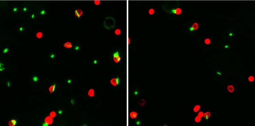
“We asked, do neutrophils also have a disdain for eating rods?” she said. “We found the complete opposite. They actually have a preference for eating rods.”
And that preference is useful for targeting neutrophils and leaving other white blood cells to do their jobs. They found that when they offered rods to different phagocytes, 80% of the neutrophils ate them, whereas only 5% to 10% of other phagocytes did.
The comparisons included macrophages, another cell that eats intruders, and dendritic cells, which capture intruders and then show the other immune cells what to look for.
The team is currently exploring whether neutrophil-distracting particles can be made from medications rather than plastic. Eniola-Adefeso is now working with the U-M Office of Technology Transfer to advance her delivery system toward clinical trials, in hopes that it may prove useful in the fight against COVID-19.
U-M has applied for patent protection and has launched a start-up company, Asalyxa.
Eniola-Adefeso is also a professor of biomedical engineering and professor of macromolecular science and engineering.
The paper is titled, “Neutrophils preferentially phagocytose elongated particles – opportunity for selective targeting in acute inflammatory diseases,” and is published in the journal Science Advances.
SARS-CoV-2, immune response and the cytokine storm.
When SARS CoV-2, an enveloped, single-stranded, positive-sense RNA virus is inhaled, it gains entry into the host cell in a two-stage process. (5)
First, the viral spike (S) protein binds to the ACE-2 receptor, located on type-II cells. This facilitates viral attachment. The S protein is then further primed for fusion and entry into the host cell by the protease, TMPRSS2.
As a result of this binding and host invasion by SARS-CoV-2 both the innate and the adaptive immune systems are activated, with the consequent release of numerous types of cytokines including IL-6. (6)
This constitutes a functional immune response.
However, in severe COVID-19 patients the immune response becomes dysregulated, with a pathological effect on both inflammation and autoimmunity. In particular, there are elevated levels of IL-6 (7, 8), which contributes to the cytokine storm, an immune system over-reaction.
IL-6 levels in COVID-19 patients have been reported to exceed 627.1pg/mL before TCZ therapy. The normal level is 5pg/mL or less.
Aside from elevated IL-6 levels, severe COVID-19 patients also have lower levels of beneficial CD4+ and CD8+ T cells, both of which are part of the adaptive (or acquired) immune system.
This effect of IL-6 confirms that the cytokine storm diminishes adaptive immunity against SARS-CoV- 19 infection, thus making the patient less likely to recover. As Liu, et al (2020) has noted, “The more serious the disease and the worse the prognosis, the lower were the T cell, CD4+ T cell, and CD8+ T cell counts on admission. Based on these findings, we believe that the CD4+ and CD8+ T cell counts in patients with COVID-19 could reflect disease severity and predict disease prognosis and are therefore good biomarkers of COVID-19 activity.”
In short, IL-6 shifts from acting to protect the host due via a regulated immune system, to becoming a threat to host survival due to dysregulated immune system.
Mechanism of the cytokine storm.
The replication and release of the virus causes inflammatory host cell death (pyroptosis). Damage- associated molecular patterns (DAMP)are recognised by alveolar macrophages which start the production of pro-inflammatory cytokines and chemokines including IL-6.
The initial action of IL-6 is beneficial to the host as it contributes to the immune response. However, as further monocytes, macrophages and T cells are attracted to the infection site, due to the increasing number of viral particles, there is an augmentation of the inflammation process.
A pro-inflammatory cytokine “feedback loop” is created.
The dysregulated consequences associated with the increase in IL-6 includes enhanced vascular endothelial permeability, resulting in increased levels of neutrophils in the lungs.
Neutrophil entry into the lungs is a defining feature of ARDS.
Neutrophils cause an increase in capillary permeability, resulting in alveolar oedema and arterial hypoxaemia, further compromising ongoing gas exchange and adding to the onset of acute respiratory distress syndrome (ARDS).
ARDS mortality corresponds to the degree of neutrophilia in the lung. There is also a diffuse thickening of the alveolar wall, further decreasing gas exchange.
Respiratory failure caused 69.5% of deaths in one Wuhan hospital cohort of 82 patients.
Another consequence of neutrophil-induced capillary permeability is that pro-inflammatory IL-6 can escape into the systemic circulation. The systemic ‘escape’ of supraphysiological levels of IL-6 is implicated in multi-organ damage, including kidney disease, cardiovascular disease (including fulminant myocarditis)(9), and hepatic failure.(10)
The systemic viral damage occurs because IL-6 receptors are ubiquitous. Foetal tissue studies show IL-6 receptors not only in the lungs, but also the eye, heart, liver, spleen, adrenal, renal and gastrointestinal tissue.
Adding to the consequences of the cytokine storm is clinical evidence from human and animal studies indicated that an “overexpression of IL-6 might be a possible mechanism favouring persistence of some viruses”, perhaps secondary to the reduced levels of CD4+ and CD8+ T cells noted earlier.
The multi-factorial interplay of immune components, cytokines and SARS-CoV-2 is well represented in Figure 1, from Pedersen et al, reproduced with permission.

SARS-CoV2, cardiovascular complications and ACE2 receptors.
Research indicates that there are several prognostic indicators for severe disease and ICU admission in COVID-19 patients. These include being elderly and having underlying comorbidities such as chronic obstructive pulmonary disease (COPD), cardiovascular disease and hypertension.
One study has reported that 21% of COVID-19 patients had hypertension. Hypertension is associated with a nearly 2.5- fold significantly increased risk of severe COVID-19 disease and a similar significantly higher risk of mortality.
Cardiovascular morbidity – characterised by tachyarrhythmias, elevated troponin, and thromboembolic events – is reported in 20% of hospitalised patients and is strongly allied with mortality risk. (11)
The elucidated explanation for the higher mortality rate in hypertensive patients is due to the destruction of the ACE2 receptors after SARS-Cov2 binding to the type II cells and subsequent cell lysis.
Importantly, ACE2 receptors are also located on the heart, gastrointestinal and kidneys at higher levels than are found in the lungs.
The physiological function of ACE2 is to convert angiotensin II to angiotensin 1-7. The ACE2- angiotensin 1-7 axis is a counterbalancing arm to the ACE1-angiotensin II arm of the renin-angiotensin system (RAS).
Angiotensin II can induce vasoconstriction and possesses proinflammatory and profibrotic capacities. In contrast, angiotensin 1-7 is antiproliferative, antiapoptotic and has vasodilating actions. It is also cardioprotective in its capacity to be anti-heart failure, antithrombotic, anti-myocardial hypertrophy, antifibrosis, antiarrhythmic and anti-atherogenic.
As can be appreciated, with the destruction of ACE2 receptors on type II cells, the delicate balance between ACE1 and ACE2 can lead to dysregulation of blood pressure and other deleterious cardiovascular sequelae.
The role and interplay between SARS-CoV2, the RAS and higher mortality rates are elegantly portrayed in Figure 2, from the paper by Guo et al (Open Access Article).

respiratory distress syndrome; AT1R, angiotensin II type 1 receptor; dotted line, speculation based on the current evidence; solid line, findings from current evidence; up arrow, promote; down arrow, inhibit.
SARS-CoV-2 and stroke in COVID-19 patients.
On April 28, 2020 the NEJM reported on five COVID-19 patients who present with large-vessel stroke.
(12) A possible explanation may involve virus induced destruction of the ACE-2 receptor and reduced AT1-7 generation, the production of superoxides causing resultant endothelial damage, and the putative impact of this cascade on the release of von Willebrand factor (vWF) and the link to clot formation and stroke. The release of vWF is also triggered by IL-6. (13-17)
Kerr (2001) reported that IL-6 promotes coagulation via an increased transcription of Factor VIII as well as causing a 4.5-fold increase in tissue factor mRNA. IL-6 also increases both platelet production and activation, and von Willebrand factor (vWF). (18) Similarly, Chin (2003) has reported that “elevated IL- 6 may contribute to the thrombotic and thromboembolic complications in acute heart failure, in a process mediated via increased TF and vWF.” (19)
Peyvandhi (2011) and Escher (2020) have concluded that VWF plays a pivotal role in primary haemostasis by facilitating platelet adhesion to damaged vascular endothelium and subsequent platelet aggregation. (20, 21) High levels of vWF, in part caused by elevated IL-6 levels, are predictive of endothelial activation, disease outcome and future prothrombotic events. (21, 22)
References:
- STATISTA. Number of coronavirus (COVID-19) cases, recoveries, and deaths among the most impacted countries worldwide as of April 24, 2020 2020. Available from: https://www.statista.com/statistics/1105235/coronavirus-2019ncov-cases-recoveries-deaths-most-affected- countries-worldwide/.
- D’Elia RV, Harrison K, Oyston PC, Lukaszewski RA, Clark GC. Targeting the “Cytokine Storm” for Therapeutic Benefit. Clinical and Vaccine Immunology [Internet]. 2013; 20(3):[319-27 pp.]. Available from: https://cvi.asm.org/content/cdli/20/3/319.full.pdf.
- Choy E, Rose-John S. Interleukin-6 as a Multifunctional Regulator: Inflammation, Immune Response, and Fibrosis. Journal of Scleroderma and Related Disorders [Internet]. 2017; 2(2_suppl):[S1-S5 pp.]. Available from: https://journals.sagepub.com/doi/abs/10.5301/jsrd.5000265.
- Tanaka T, Narazaki M, Kishimoto T. IL-6 in inflammation, immunity, and disease. Cold Spring Harb Perspect Biol [Internet]. 2014; 6(10):[a016295-a pp.]. Available from: https://pubmed.ncbi.nlm.nih.gov/25190079
- Yang PW, Xiliang. COVID-19: a new challenge for human beings. Cellular & Molecular Immunology [Internet]. 2020 2020/03/31. Available from: https://doi.org/10.1038/s41423-020-0407-x.
- Zhang C, Wu Z, Li J-W, Zhao H, Wang G-Q. The cytokine release syndrome (CRS) of severe COVID-19 and Interleukin-6 receptor (IL-6R) antagonist Tocilizumab may be the key to reduce the mortality. International journal of antimicrobial agents [Internet]. 2020:[105954- pp.]. Available from: https://pubmed.ncbi.nlm.nih.gov/32234467
- Pedersen SF, Ho Y-C. SARS-CoV-2: a storm is raging. The Journal of clinical investigation [Internet]. 2020 04/13/; 130(5). Available from: https://doi.org/10.1172/JCI137647.
- Michot J-MA, L. Tocilizumab, an anti-IL6 receptor antibody, to treat Covid-19-related respiratory failure – a case report2020 24/04/2020. Available from: https://www.annalsofoncology.org/article/S0923- 7534(20)36387-0/pdf.
- Ruan Q, Yang K, Wang W, Jiang L, Song J. Clinical predictors of mortality due to COVID-19 based on an analysis of data of 150 patients from Wuhan, China. Intensive care medicine [Internet]. 2020 Mar 3 PMC7080116]. Available from: https://www.ncbi.nlm.nih.gov/pmc/articles/PMC7080116/pdf/134_2020_Article_5991.pdf.
- Tay MZ, Poh CM, Rénia L, MacAry PA, Ng LFP. The trinity of COVID-19: immunity, inflammation and intervention. Nature Reviews Immunology [Internet]. 2020 2020/04/28. Available from: https://doi.org/10.1038/s41577-020-0311-8.
- Felsenstein S, Herbert JA, McNamara PS, Hedrich CM. COVID-19: Immunology and treatment options. Clin Immunol [Internet]. 2020; 215:[108448- pp.]. Available from: https://pubmed.ncbi.nlm.nih.gov/32353634
- Oxley TJ, Mocco J, Majidi S, Kellner CP, Shoirah H, Singh IP, et al. Large-Vessel Stroke as a Presenting Feature of Covid-19 in the Young. New England Journal of Medicine [Internet]. 2020:[e60 p.]. Available from: https://www.nejm.org/doi/full/10.1056/NEJMc2009787.
- Zhang H, Penninger JM, Li Y, Zhong N, Slutsky AS. Angiotensin-converting enzyme 2 (ACE2) as a SARS- CoV-2 receptor: molecular mechanisms and potential therapeutic target. Intensive care medicine [Internet]. 2020 Apr PMC7079879]; 46(4):[586-90 pp.]. Available from: https://www.ncbi.nlm.nih.gov/pubmed/32125455.
Patel VB, Zhong J-C, Grant MB, Oudit GY. Role of the ACE2/Angiotensin 1-7 Axis of the Renin- Angiotensin System in Heart Failure. Circulation research [Internet]. 2016; 118(8):[1313-26 pp.]. Available from: http://search.ebscohost.com/login.aspx?direct=true&db=mdc&AN=27081112&site=ehost- live&scope=site.
Panday A, Sahoo MK, Osorio D, Batra S. NADPH oxidases: an overview from structure to innate immunity-associated pathologies. Cell Mol Immunol [Internet]. 2015 Jan PMC4654378]; 12(1):[5-23 pp.]. Available from: https://www.ncbi.nlm.nih.gov/pubmed/?term=panday+NADPH+oxidases+2015.
- Loscalzo J. Oxidative stress in endothelial cell dysfunction and thrombosis. Pathophysiology of haemostasis and thrombosis [Internet]. 2002 Sep-Dec; 32(5-6):[359-60 pp.]. Available from: https://www.karger.com/Article/PDF/73600.
- Welch WJ. Angiotensin II-dependent superoxide: effects on hypertension and vascular dysfunction. Hypertension [Internet]. 2008; 52(1):[51-6 pp.]. Available from: https://pubmed.ncbi.nlm.nih.gov/18474831
- Kerr R, Stirling D, Ludlam CA. Interleukin 6 and Haemostasis. British journal of haematology [Internet]. 2001; 115(1):[3-12 pp.]. Available from: https://onlinelibrary.wiley.com/doi/abs/10.1046/j.1365- 2141.2001.03061.x.
- Chin BS, Conway DS, Chung NA, Blann AD, Gibbs CR, Lip GY. Interleukin-6, tissue factor and von Willebrand factor in acute decompensated heart failure: relationship to treatment and prognosis. Blood coagulation & fibrinolysis : an international journal in haemostasis and thrombosis [Internet]. 2003 Sep; 14(6):[515-21 pp.].
- Peyvandi F, Garagiola I, Baronciani L. Role of von Willebrand factor in the haemostasis. Blood Transfus [Internet]. 2011; 9 Suppl 2(Suppl 2):[s3-s8 pp.]. Available from: https://pubmed.ncbi.nlm.nih.gov/21839029
- Escher R, Breakey N, Lämmle B. Severe COVID-19 infection associated with endothelial activation. Thrombosis research [Internet]. 2020; 190:[62- pp.]. Available from: https://www.ncbi.nlm.nih.gov/pmc/articles/PMC7156948/.
- Luiza Rusu LM, RD. Endothelial Cell von Willebrand Factor Secretion in Health and Cardiovascular Disease 2017 09/05/2020]. Available from: DOI: 10.5772/intechopen.74029
- Jaimes JA, André NM, Chappie JS, Millet JK, Whittaker GR. Phylogenetic Analysis and Structural Modeling of SARS-CoV-2 Spike Protein Reveals an Evolutionary Distinct and Proteolytically Sensitive Activation Loop. Journal of Molecular Biology [Internet]. 2020 2020/04/19/. Available from: http://www.sciencedirect.com/science/article/pii/S0022283620302874.
- Sun A, Chia JS, Chang YF, Chiang CP. Serum interleukin-6 level is a useful marker in evaluating therapeutic effects of levamisole and Chinese medicinal herbs on patients with oral lichen planus. Journal of oral pathology & medicine : official publication of the International Association of Oral Pathologists and the American Academy of Oral Pathology [Internet]. 2002 Apr; 31(4):[196-203 pp.]. Available from: https://www.ncbi.nlm.nih.gov/pubmed/12076322.
More information: “Neutrophils preferentially phagocytose elongated particles—Opportunity for selective targeting in acute inflammatory diseases” Science Advances (2020). advances.sciencemag.org/content/6/24/eaba1474


