


 Extract (6-MSITC) in Healthy Older Adults")
: An In-Depth Exploration into its Thermogenic Role and Social Significance")


New research by neuroscientists at the University of Pittsburgh and University of California San Francisco (UCSF) revealed that a simple, earbud-like device developed at UCSF that imperceptibly stimulates a key nerve leading to the brain could significantly improve the wearer’s ability to learn the sounds of a new language.
This device may have wide-ranging applications for boosting other kinds of learning as well.
Mandarin Chinese is considered one of the hardest languages for native English speakers to learn, in part because the language – like many others around the world – uses distinctive changes in pitch, called “tones,” to change the meaning of words that otherwise sound the same.
In the new study, published today in npj Science of Learning, researchers significantly improved the ability of native English speakers to distinguish between Mandarin tones by using precisely timed, non-invasive stimulation of the vagus nerve – the longest of the 12 cranial nerves that connect the brain to the rest of the body.
What’s more, vagus nerve stimulation allowed research participants to pick up some Mandarin tones twice as quickly.
“Showing that non-invasive peripheral nerve stimulation can make language learning easier potentially opens the door to improving cognitive performance across a wide range of domains,” said lead author Fernando Llanos, Ph.D., a postdoctoral researcher in Pitt’s Sound Brain Lab.
“This is one of the first demonstrations that non-invasive vagus nerve stimulation can enhance a complex cognitive skill like language learning in healthy people,” said Matthew Leonard, Ph.D., an assistant professor, Department of Neurological Surgery, UCSF Weill Institute for Neurosciences, whose team developed the nerve stimulation device.
Leonard is a senior author of the new study, alongside Bharath Chandrasekaran, Ph.D., professor and vice chair of research, Department of Communication Science and Disorders, Pitt School of Health and Rehabilitation Sciences, and director of the Sound Brain Lab.
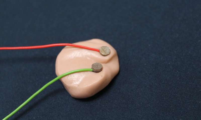
Researchers used a non-invasive technique called transcutaneous vagus nerve stimulation (tVNS), in which a small stimulator is placed in the outer ear and can activate the vagus nerve using unnoticeable electrical pulses to stimulate one of the nerve’s nearby branches.
For their study, the researchers recruited 36 native English-speaking adults and trained them to identify the four tones of Mandarin Chinese in examples of natural speech, using a set of tasks developed in the Sound Brain Lab to study the neurobiology of language learning.

Participants who received imperceptible tVNS paired with two Mandarin tones that are typically easier for English speakers to tell apart showed quick improvements in learning to distinguish these tones.
By the end of the training, those participants were 13% better on average at classifying tones and reached peak performance twice as quickly as control participants who wore the tVNS device but never received stimulation.
“There’s a general feeling that people can’t learn the sound patterns of a new language in adulthood, but our work historically has shown that’s not true for everyone,” Chandrasekaran said. “
In this study, we are seeing that tVNS reduces those individual differences more than any other intervention I’ve seen.”
“This approach may be leveling the playing field of natural variability in language learning ability,” added Leonard.
“In general, people tend to get discouraged by how hard language learning can be, but if you could give someone 13% to 15% better results after their first session, maybe they’d be more likely to want to continue.”
The researchers now are testing whether longer training sessions with tVNS can impact participants’ ability to learn to discriminate two tones that are harder for English speakers to differentiate, which was not significantly improved in the current study.
Stimulation of the vagus nerve has been used to treat epilepsy for decades and has recently been linked to benefits for a wide range of issues ranging from depression to inflammatory disease, though exactly how these benefits are conferred remains unclear.
But most of these findings have used invasive forms of stimulation involving an impulse generator implanted in the chest.
By contrast, the ability to evoke significant boosts to learning using simple, non-invasive vagus nerve stimulation could lead to significantly cheaper and safer clinical and commercial applications.
The researchers suspect tVNS boosts learning by broadly enhancing neurotransmitter signaling across wide swaths of the brain to temporarily boost attention to the auditory stimulus being presented and promote long-term learning, though more research is needed to verify this mechanism.
“We’re showing robust learning effects in a completely non-invasive and safe way, which potentially makes the technology scalable to a broader array of consumer and medical applications, such as rehabilitation after stroke,” Chandrasekaran said.
“Our next step is to understand the underlying neural mechanism and establish the ideal set of stimulation parameters that could maximize brain plasticity. We view tVNS as a potent tool that could enhance rehabilitation in individuals with brain damage.”
Neuromodulation-based approached to enhancing foreign language learning and skill training outcomes
Studies examining the neurobiology of language learning have shown diverse brain regions are involved.
Functional plasticity in these brain areas appears to be a key determinant in the ability of individuals to learn languages from early childhood through adulthood.
From a classical perspective, the activity and plasticity of two well characterized language-related brain regions have long been known to play major roles in our ability to learn, communicate with, and comprehend spoken languages.
Broca’s area is necessary for the planning and execution of speech while Wernicke’s area is required for the analysis and identification of speech [1, 2].
Stemming from several decades of neurophysiological and neuroimaging studies however, we have expanded our understanding of language learning to include many sparsely distributed anatomical regions.
For example the perisylvian cortex, superior and middle temporal gyri, the lingual and fusiform gyri, inferior temporal cortex, dorsolateral prefrontal cortex, insula, central operculum, anterior cingulate, and basal ganglia have been shown to play roles in learning, listening, speaking, repeating, and comprehension of spoken language [1, 2].
Further, primary sensory brain regions like the auditory cortex and memory-encoding or information consolidation regions like the hippocampus are also required for the acquisition and proficient use of language.
Therefore neuromodulation-based strategies designed to enhance language learning might benefit from influencing the activity and plasticity of multiple linguistically-relevant anatomical loci, spatially distributed cognitive control networks, and sensory processing regions in a simultaneous or coordinated manner.
Several methods of enhancing language learning have been described in the literature. Major advances have been made over the past decade in the design and use of autonomous learning methods, such as those currently incorporated in mobile-based language training applications or computer-assisted language learning programs [3–5].
More recent advances in virtually immersive or virtual reality (VR) technologies have also proven useful for enhancing foreign language learning [6, 7]. While these VR and computer-assisted cognitive training methods can enhance language acquisition by facilitating natural brain activity patterns arising from repeated training sessions or sensory stimulation, they do not directly augment brain activity or plasticity to facilitate learning per se.
Sleep has also been shown to play a critical role in the consolidation learned information including spoken-language learning [8]. Recent efforts to influence the consolidation phases of language learning during sleep have also shown promise by using targeted memory reactivation (TMR).
These TMR investigations demonstrated word cuing during certain stages of sleep can enhance vocabulary learning [9–11]. These TMR methods require the implementation of electroencephalography (EEG) to identify certain stages of sleep, such as slow wave sleep during which sensory information or linguistic cues are presented to enhance language training outcomes.
Using EEG during sleep poses several technical challenges that can interfere with broad adoption for enhancing language learning.
Other noninvasive neuromodulation methods, which have been developed to directly augment brain activity and plasticity have also been used to enhance language learning.
Transcranial magnetic stimulation (TMS) and transcranial direct current stimulation (tDCS) are two forms of noninvasive neuromodulation that can target restricted regions of cortex to influence brain activity and plasticity.
Studies have demonstrated that TMS and tDCS delivered to classical language brain regions like Wernicke’s and Broca’s areas can enhance plasticity and language recovery in patients suffering from post-stroke aphasias [12].
In non-aphasic, healthy volunteers tDCS of Wernicke’s area has been shown to improve verbal memory, enhance word retrieval, and reduce interference during word learning [13, 14].
Also in healthy adults tDCS has been shown to enhance language learning when applied to the posterior part of the left peri-sylvian area [15], enhance language performance when delivered to the left prefrontal cortex [16], improve word retrieval when applied to the primary motor cortex of older adults [17], and to facilitate the learning and maintenance of a novel vocabulary when administered acutely to the temporo-parietal junction on several consecutive days [18].
Despite the success of these methods they are restricted by their coarse targeting abilities, can only modulate one cortical network at a time, and are limited to modulating top-down cortical processes since they are incapable of reaching deeper brain structures.
With the major objective of developing Targeted Neuroplasticity Training (TNT) methods for accelerating foreign language learning, we began to explore the utility of cranial nerve neuromodulation modalities, such as vagal nerve stimulation (VNS).
Stimulation of the cervical and auricular branches of the vagus nerve has been repeatedly demonstrated to induce short-term and long-term brain plasticity [19].
It has also been shown to modulate brain circuits and trigger signaling consequences supporting mechanisms of action for the observed plasticity [20–23].
Superficial branches of the vagus in particular the auricular branch of the vagus nerve (ABVN) are located in anatomical regions such that achieving functional electrical coupling to them can be achieved using a number of medical electrode designs including conventional transdermal electrical nerve stimulation (TENS) approaches.
Vagus Nerve Stimulation for Enhancing Human Brain Plasticity
Electrical stimulation of vagal afferents in several animal species, including humans, has been shown to exert prominent bottom-up actions on key neuromodulatory brain circuits, such as the locus coeruleus (LC) and dorsal raphe (DR), as well as powerful endogenous neurotransmitters like norepinephrine (NE) and serotonin (5-HT) that are known to regulate arousal, attention, and neurophysiological/behavioral plasticity (Figure 1; [20–31].
Further, data from several studies has shown that electrical modulation of vagal afferents, including the ABVN, in healthy humans can safely produce biochemical, behavioral, and neurophysiological effects that are consistent with an enhancement of learning or skill training [32–34].
Based on this collective evidence we hypothesized that taVNS may represent a human factor optimized approach to achieving TNT enhancement of language learning.

Modulation of peripheral nerves of the external ear for Targeted Neuroplasticity Training. Transcutaneous electrical stimulation of the auricular branch of the vagus and other nerves influence ascending brain activity and arousal systems to enhance skilled training and learning.
One of the major hurdles for developing VNS methods and systems intended to optimize skill training and learning will be to identify the optimal stimulation parameters required to drive acute neural plasticity.
While the VNS parameters used across investigations are somewhat uniform, there are not standardized VNS protocols. Systematic studies have not been conducted to identify the best VNS parameters for optimizing neuromodulator signaling or plasticity in the healthy adult brain.
Many of the VNS parameters that have been used to date were originally identified and adopted for their ability to desynchronize or interfere with aberrant neural activity like that, which occurs in diseased brain states, such as epilepsy, tinnitus, and depression.
In fact, the VNS studies that have shown effects on plasticity have largely borrowed their stimulation devices and parameters from the clinical designs and practices [21,27,32–35].
This poses somewhat of an issue when developing a VNS system for enhancing normal plasticity related to training and learning. It is simply not known whether clinically identified and validated neurostimulation parameters are ideally suited for VNS applications where optimizing normal brain function is the desired outcome.
Therefore, a significant amount of our performance effort has been devoted to investigating how different VNS strategies affect sensory or auditory plasticity and autonomic arousal.
Targeting Auricular Branches of the Human Vagus Nerve
Anatomical and functional studies have shown the afferents of the AVBN innervate the superficial regions of the auricle specifically along the external meatus and concha (Figures 1 and 2A).
For example, a study investigating the anatomical evidence for the involvement of the AVBN in Arnold’s cough-reflex showed branches of the AVBN project to the meatus in cadaver microdissection and high-resolution CT imaging studies [36].
Another eloquent histological study conducted on human tissues presented robust histological and immunocytological evidence for the presence of vagal fibers lining the skin outside of the cartilage of the meatus [37].
The presence of fewer fibers was observed in the skin of the concha and moreover those nerves were deeply embedded in the cartilage [37]. Based on this evidence we hypothesized the auditory meatus to be an idealized target for taVNS since there is a higher density of nerve fibers in this location and since cartilage will not interfere with the delivery of pulsed currents to the nerves as much as in other external ear regions.
Therefore we engineered, optimized, designed and fabricated biocompatible, hydrogel-based earbud electrodes designed for wearing in the auditory meatus to gain preliminary insights into the safety and efficacy of this method for enhancing sensory processing and brain plasticity.
We specifically investigated how varied taVNS stimulus protocols delivered through earbud electrodes influence the safety, tolerability, acute auditory plasticity, and autonomic physiology of healthy human volunteers.
Human Dimensions & Real-world Considerations for Auricular Vagus Nerve Stimulation Methods
Transdermal auricular vagal nerve stimulation (taVNS) has been shown to be a largely safe and effective method of regulating ascending neuromodulatory brainstem circuits, as well as higher cortical regions in clinical populations.
Evidence has suggested this safety extends into a healthy population since VNS has shown be safe and effective for modulating brain activity in healthy adults in many studies now.
A potential hurdle to using previously methods described is that they use misguided targeting approaches and they have implemented poor industrial designs that properly account for real world human factors, such as comfort or ability to wear for extended durations.
Many taVNS reports to date have targeted the tragus, for example using crude methods with electrode clips [38]. It remains unclear why other than for convenience or lack of knowledge with respect to functional vagal nerve neuroanatomy.
This method is problematic for extended wear due to electrode movement and potential user pain or discomfort.
Electrode clips can become quite uncomfortable due to mechanical pinching.
Further some of these clip electrodes are made from a high impedance rubber or carbon that is not ideal for noninvasive, transcutaneous electrical nerve modulation.
Other fairly coarse approaches have implemented small (1-2 mm) stainless-steel ball electrodes positioned at two or more locations in the concha and external ear [39], which can result in discomfort due to high current densities at the electrode-skin interface.
In our investigations, we aimed to ensure wear comfort and comfort during electrical stimulation treatments were optimized as not to distract users or induce pain that may cause significant confounds when trying to enhance brain plasticity or optimize human performance.
Many other reports exist in the field that have implemented poorly designed approaches to electrically interfacing with the skin of the external ear.
For instance, Keute and colleagues (2019) used paper or cloth-backed backed electrodes with electrode coupling paste to position small (4 × 4 mm) electrodes in the concha.
This again can produce locally high current densities due to the small electrode size used. Further it is a methodological approach subject to numerous confounds and inconsistencies due to unavoidable and significant fluctuations in electrode-skin impedances that will occur with motion or very small movements.
Moreover, the electrode materials used were not ideally suited for taVNS.
Perhaps most concerning is that the implementation of the above discussed methods can produce unreliable results, cloud the field with murky data, impede future progress towards the development of real world applications, and can influence public perception and funding initiatives.
Therefore, in our investigations, we aimed to utilize Best Practices from state-of-the-art peripheral nerve stimulation methods, as well as modern electrode design and engineering principles.
We specifically ensured individualized and proper electrode fits to minimize mechanical movement and electrical impedance fluctuations while maximizing wear comfort and comfort during electrical stimulation treatments.
These practices are critical because the methods should not to distract users or induce any pain/discomfort that may cause significant confounds when trying to enhance brain plasticity, modulate arousal and attention, or optimize human performance.

Overview of transdermal auricular vagal nerve stimulation approaches. A) Anatomical illustration showing several branches of cranial nerves (CN V auriculotemporal branch of the trigeminal nerve, CN VII facial nerve, and CN X auricular branch of the vagus nerve = AVBN) and cervical nerves (C2 and C3; lessor occipital nerve = LON and great auricular nerve = GAN) that innervate the external ear. We targeted the ABVN using custom (B) or semi-custom/modified (C) ear bud taVNS electrodes inserted into the acoustic meautus. D) Biphasic, charge-balanced, pulsed electrical currents having varied pulse frequencies and pulse widths (Pw) with an interphase gap (Pg) of 50 microseconds were delivered at different peak current intensities (Ipeak) to the left or both ears through taVNS electrodes after (baseline) and before passive auditory listening tasks while EEG and physiological data were recorded.
References
1. Shtyrov Y: Neural bases of rapid word learning. The Neuroscientist: A Review Journal Bringing Neurobiology, Neurology and Psychiatry 2012, 18:312–319.
2. Neville HJ, Bavelier D: Neural organization and plasticity of language. Current Opinion in Neurobiology 1998, 8:254–258.
3. Godwin-Jones R: Mobile Apps for Lanugage Learning. Language Learning & Technology 2011, 15:2–11.
4.Stevens C, Fanning J, Coch D, Sanders L, Neville H: Neural mechanisms of selective auditory attention are enhanced by computerized training: Electrophysiological evidence from language-impaired and typically developing children. Brain Research 2008, 1205:55–69.
5. Rosell-Aguilar F: Autonomous language learning through a mobile application: a user evaluation of the busuu app. Computer Assisted Language Learning 2018, 31:854–881.
6. Shih Y-C, Yang M-T: A Collaborative Virtual Environment for Situated Language Learning Using VEC3D. Journal of Educational Technology & Society 2008, 11:56–68.
7. Schwienhorst K: Why Virtual, Why Environments? Implementing Virtual Reality Concepts in Computer-Assisted Language Learning. Simulation & Gaming 2002, 33:196–209.
8. Fenn KM, Nusbaum HC, Margoliash D: Consolidation during sleep of perceptual learning of spoken language. Nature 2003, 425:614–616.
9. Schreiner T, Rasch B: Boosting Vocabulary Learning by Verbal Cueing During Sleep. Cerebral Cortex (New York, N.Y.: 1991) 2015, 25:4169–4179.
10.Schreiner T, Rasch B: The beneficial role of memory reactivation for language learning during sleep: A review. Brain and Language 2017, 167:94–105.
11. Züst MA, Ruch S, Wiest R, Henke K: Implicit Vocabulary Learning during Sleep Is Bound to Slow-Wave Peaks. Current Biology.
12. Hamilton RH, Chrysikou EG, Coslett B: Mechanisms of aphasia recovery after stroke and the role of noninvasive brain stimulation. Brain and Language 2011, 118:40–50.
13. Fiori V, Coccia M, Marinelli CV, Vecchi V, Bonifazi S, Ceravolo MG, Provinciali L, Tomaiuolo F, Marangolo P: Transcranial direct current stimulation improves word retrieval in healthy and nonfluent aphasic subjects. Journal of Cognitive Neuroscience 2011, 23:2309–2323.
14. Rivera-Urbina GN, Mendez Joya MF, Nitsche MA, Molero-Chamizo A: Anodal tDCS over Wernicke’s area improves verbal memory and prevents the interference effect during words learning. Neuropsychology 2019, 33:263–274.
15. Flöel A, Rösser N, Michka O, Knecht S, Breitenstein C: Noninvasive Brain Stimulation Improves Language Learning. Journal of Cognitive Neuroscience 2008, 20:1415–1422.
16. Hussey EK, Ward N, Christianson K, Kramer AF: Language and Memory Improvements following tDCS of Left Lateral Prefrontal Cortex. PLoS ONE 2015, 10.
17. Meinzer M, Lindenberg R, Sieg MM, Nachtigall L, Ulm L, Flöel A: Transcranial direct current stimulation of the primary motor cortex improves word-retrieval in older adults. Frontiers in Aging Neuroscience 2014, 6:253.
18. Meinzer M, Jähnigen S, Copland DA, Darkow R, Grittner U, Avirame K, Rodriguez AD, Lindenberg R, Flöel A: Transcranial direct current stimulation over multiple days improves learning and maintenance of a novel vocabulary. Cortex; a Journal Devoted to the Study of the Nervous System and Behavior 2014, 50:137–147.
19. Ben-Menachem E, Revesz D, Simon BJ, Silberstein S: Surgically implanted and non-invasive vagus nerve stimulation: a review of efficacy, safety and tolerability. Eur J Neurol 2015, 22:1260–1268.
20. Fang J, Rong P, Hong Y, Fan Y, Liu J, Wang H, Zhang G, Chen X, Shi S, Wang L, et al: Transcutaneous Vagus Nerve Stimulation Modulates Default Mode Network in Major Depressive Disorder. Biol Psychiatry 2016, 79:266–273.
21. Frangos E, Ellrich J, Komisaruk BR: Non-invasive Access to the Vagus Nerve Central Projections via Electrical Stimulation of the External Ear: fMRI Evidence in Humans. Brain stimulation 2015, 8:624–636.
22.Krahl SE, Clark KB: Vagus nerve stimulation for epilepsy: A review of central mechanisms. Surgical Neurology International 2012, 3:S255–S259.
23. Kraus T, Hösl K, Kiess O, Schanze A, Kornhuber J, Forster C: BOLD fMRI deactivation of limbic and temporal brain structures and mood enhancing effect by transcutaneous vagus nerve stimulation. Journal of Neural Transmission 2007, 114:1485–1493.
24.George MS, Aston-Jones G: Noninvasive techniques for probing neurocircuitry and treating illness: vagus nerve stimulation (VNS), transcranial magnetic stimulation (TMS) and transcranial direct current stimulation (tDCS). Neuropsychopharmacology 2010, 35:301–316.
25.Shiozawa P, da Silva ME, de Carvalho TC, Cordeiro Q, Brunoni AR, Fregni F: Transcutaneous vagus and trigeminal nerve stimulation for neuropsychiatric disorders: a systematic review. Arq Neuropsiquiatr 2014, 72:542–547.
26.Raedt R, Clinckers R, Mollet L, Vonck K, El Tahry R, Wyckhuys T, De Herdt V, Carrette E, Wadman W, Michotte Y, et al: Increased hippocampal noradrenaline is a biomarker for efficacy of vagus nerve stimulation in a limbic seizure model. Journal of Neurochemistry 2011, 117:461–469.
27. Borland MS, Vrana WA, Moreno NA, Fogarty EA, Buell EP, Sharma P, Engineer CT, Kilgard MP: Cortical Map Plasticity as a Function of Vagus Nerve Stimulation Intensity. Brain Stimulation: Basic, Translational, and Clinical Research in Neuromodulation 2016, 9:117–123.
28.Manta S, Dong J, Debonnel G, Blier P: Enhancement of the function of rat serotonin and norepinephrine neurons by sustained vagus nerve stimulation. J Psychiatry Neurosci 2009, 34:272–280.
29.Manta S, Dong J Fau – Debonnel G, Debonnel G Fau – Blier P, Blier P: Optimization of vagus nerve stimulation parameters using the firing activity of serotonin neurons in the rat dorsal raphe. 2009.
30.Kraus T, Kiess O, Hösl K, Terekhin P, Kornhuber J, Forster C: CNS BOLD fMRI Effects of Sham-Controlled Transcutaneous Electrical Nerve Stimulation in the Left Outer Auditory Canal – A Pilot Study. Brain Stimulation: Basic, Translational, and Clinical Research in Neuromodulation 2013, 6:798–804.
31. Hays SA, Rennaker RL, Kilgard MP: Targeting plasticity with vagus nerve stimulation to treat neurological disease. Prog Brain Res 2013, 207:275–299.
32. Jacobs HIL, Riphagen JM, Razat CM, Wiese S, Sack AT: Transcutaneous vagus nerve stimulation boosts associative memory in older individuals. Neurobiology of Aging 2015, 36:1860–1867.
33.Sellaro R, van Leusden JW, Tona KD, Verkuil B, Nieuwenhuis S, Colzato LS: Transcutaneous Vagus Nerve Stimulation Enhances Post-error Slowing. J Cogn Neurosci 2015, 27:2126–2132.
34. Steenbergen L, Sellaro R, Stock A-K, Verkuil B, Beste C, Colzato LS: Transcutaneous vagus nerve stimulation (tVNS) enhances response selection during action cascading processes. European Neuropsychopharmacology 2015, 25:773–778.
35. Engineer ND, Riley JR, Seale JD, Vrana WA, Shetake JA, Sudanagunta SP, Borland MS, Kilgard MP: Reversing pathological neural activity using targeted plasticity. Nature 2011, 470:101–104.
36. Tekdemir I, Aslan A, Elhan A: A clinico-anatomic study of the auricular branch of the vagus nerve and Arnold’s ear-cough reflex. Surgical and radiologic anatomy: SRA 1998, 20:253–257.
37. Bermejo P, Lopez M, Larraya I, Chamorro J, Cobo JL, Ordonez S, Vega JA: Innervation of the Human Cavum Conchae and Auditory Canal: Anatomical Basis for Transcutaneous Auricular Nerve Stimulation. Biomed Res Int 2017, 2017:7830919.
38. Au – Badran BW, Au – Yu AB, Au – Adair D, Au – Mappin G, Au – DeVries WH, Au – Jenkins DD, Au – George MS, Au – Bikson M: Laboratory Administration of Transcutaneous Auricular Vagus Nerve Stimulation (taVNS): Technique, Targeting, and Considerations. JoVE 2019:e58984.
39. Warren CM, Tona KD, Ouwerkerk L, van Paridon J, Poletiek F, van Steenbergen H, Bosch JA, Nieuwenhuis S: The neuromodulatory and hormonal effects of transcutaneous vagus nerve stimulation as evidenced by salivary alpha amylase, salivary cortisol, pupil diameter, and the P3 event-related potential. Brain Stimulation: Basic, Translational, and Clinical Research in Neuromodulation 2019, 12:635–642.
40. Badran BW, Yu AB, Adair D, Mappin G, DeVries WH, Jenkins DD, George MS, Bikson M: Laboratory Administration of Transcutaneous Auricular Vagus Nerve Stimulation (taVNS): Technique, Targeting, and Considerations. LID – 10.3791/58984 [doi]. Journal of Visualized Experiments (JoVE) 2019, 7.
41. Jongkees BJ, Immink MA, Finisguerra A, Colzato LS: Transcutaneous Vagus Nerve Stimulation (tVNS) Enhances Response Selection During Sequential Action.
42. Geller J, Landrigan, J.-F., & Mirman, D.: A Pupillometric Examination of Cognitive Control in Taxonomic and Thematic Semantic Memory. Journal of Cognition 2019, 2.
43. Kret ME, Sjak-Shie EE: Preprocessing pupil size data: Guidelines and code. Behavior Research Methods 2019, 51:1336–1342.
More information: npj Science of Learning, DOI: 10.1038/s41539-020-0070-0


