


 Extract (6-MSITC) in Healthy Older Adults")
: An In-Depth Exploration into its Thermogenic Role and Social Significance")


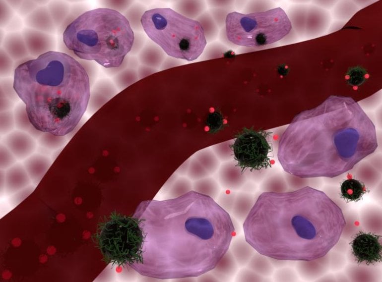
The study, published in the Journal of Materials Chemistry B, found that “loading” a chemotherapy drug on to tiny magnetic particles that can heat up the cancer cells at the same time as delivering the drug to them was up to 34% more effective at destroying the cancer cells than the chemotherapy drug without added heat.
The magnetic iron oxide nanoparticles that carry the chemotherapy drug shed heat when exposed to an alternating magnetic field.
This means that, once the nanoparticles have accumulated in the tumour area, an alternating magnetic field can be applied from outside the body, allowing heat and chemotherapy to be delivered simultaneously.
The effects of the two treatments were synergistic – that is, each treatment enhanced the effectiveness of the other, meaning they were more potent when combined than when separate. The study was carried out on cells in a lab and further research is needed ahead of clinical trials involving patients.
Senior author Professor Nguyen T. K. Thanh (Biophysics Group, UCL Physics & Astronomy) said: “Our study shows the enormous potential of combining chemotherapy with heat treatment delivered via magnetic nanoparticles.
“While this combination of therapy is already approved for the treatment of fast-growing glioblastomas, our results suggest it has potential to be used more widely as a broad anti-cancer therapy.
“This therapy also has potential to reduce the side effects of chemotherapy, by ensuring it is more highly targeted on cancer cells rather than healthy tissue. This needs to be explored in further pre-clinical tests.”
In the study, researchers combined the magnetic nanoparticles with a commonly used chemotherapy drug, doxorubicin, and compared the effects of this composite in various scenarios on human breast cancer cells, glioblastoma (brain cancer) cells, and mouse prostate cancer cells.
In the most successful scenario, they found that heat and doxorubicin together killed 98% of brain cancer cells after 48 hours, when doxorubicin without heat killed 73%. Meanwhile, for the breast cancer cells, 89% were killed by heat and doxorubicin together, while 77% were killed after 48 hours by doxorubicin alone.
Cancer cells are more susceptible to heat than healthy cells – they undergo a slow death (apoptosis) once the temperature reaches 42 degrees Celsius, whereas healthy cells are able to withstand temperatures up to 45 degrees Celsius.
The researchers found that heating cancer cells by only a few degrees, to 40 degrees Celsius, enhanced the effectiveness of the chemotherapy, meaning the treatment could be effective with lower doses of nanoparticles.
They found the combination of therapies was most effective when the nanoparticles were absorbed, or internalized, by the cancer cells, but they found the chemotherapy was also enhanced when the nanoparticles shed heat while remaining outside the cancer cells (which would be an easier form of treatment to deliver).
However, the effects at lower temperatures only occurred when the iron oxide nanoparticles were internalized or tightly deposited on to the surface of the cancer cells.
The nanoparticles also have a polymer coating that prevents the chemotherapy drug from leaching out into healthy tissue.
The coating is heat and pH-sensitive, and is designed to release the drug when temperature rises and the nanoparticles are internalized within tiny pockets in cells called “lysosomes”, which have a lower pH than the rest of the cell medium.
This intracellular delivery of the drug was particularly effective for the mouse prostate cancer cells, which showed superior and synergetic cell death effect, especially when the temperature reached 42°C.
Co-author Dr Olivier Sandre, of the University of Bordeaux, said: “Since heat can be generated through the alternating magnetic field, the release of the drug can be highly localised to cancer cells, potentially reducing side effects.”
Cancer is a class of diseases marked by the malignant proliferation of tumor cells. Despite of the huge advancements achieved during the past decades, cancer still a serious public health issue, which leading the main deaths around the world every year 1.
With the development of nanotechnology, multifunctional nanoparticle-based systems have been emerged as a new approach for efficient cancer diagnosis and treatment due to their inherent advantages in overcoming the deficiencies compared to the traditional cancer diagnostic and therapeutic techniques, such as low efficacy, drug resistance or other side effects 2-5.
Currently, various multifunctional nanoparticle-based systems integrating nanotechnology and molecular biology together have been developed as powerful tools for cancer early-stage diagnosis, real-time imaging and precise therapy 6-10. The integration of single functional component within a multifunctional therapeutic system has been demonstrated as one effective treatment approach for fighting the cancer 11-13.
The ideal nanoparticle-based diagnosis and treatment systems should not only contain the unique physical, chemical and medical properties of nanoparticles, but also be smart carriers to effectively deliver anticancer agents, thus realizing multi-modal imaging and combined therapeutic effects 14-17. Moreover, the diverse nanostructures and surface modification of nanomaterials should endow themselves with the tumor-targeting ability by EPR effect or other interactions affiliated to the features of tumor tissues 18-21.
Magnetic iron oxide nanoparticles (Fe3O4 NPs) are one representative candidate of multifunctional nanomaterials with increasing utilization in many biomedical fields, including magnetic resonance imaging (MRI), biological catalysis, magnetic hyperthermia, magnetic targeting, magnetic separation, photo-responsive therapy and drug-delivery, and currently have been widely used in tumor diagnosis and treatment 22-28.
Meanwhile, according to the specific treatment demand and characteristics of the tumor microenvironment, decoration of the Fe3O4 NPs with different functionalization factors can form the multifunctional iron oxide nanoparticles to achieve better therapeutic effects 29-31.
Compared to other imaging modalities in clinic, MRI exhibits high spatial resolution with rapid in vivo image acquisition 32, 33. To improve its sensitivity, MRI contrast agents are usually used, which can be divided into two types, namely T1 and T2 agents depending on their unique effects to alter the longitudinal or transverse relaxation time of water protons 34-36.
Compared to the widely used T1-weighted MRI agents of gadolinium (Gd)-based contrast agents with shortcoming of unpredictable renal toxicity and blood circulation time, Fe3O4 NPs are contrast agents that can be used for either T1 or T2-weighted imaging with better biosecurity 13, 37-39.
In particular, Fe3O4 NPs is a typical T2 contrast agent, when the size of Fe3O4 less than 5 nm, the decreased magnetic moment will strongly suppressed T2 effect, thus they can also be used for T1-weighted MRI imaging, which provides a possibility to prepare responsive T2-T1 switching MRI contrast agents 40, 41.
Due to the excellent thermal effect under the oscillating magnetic field, magnetic Fe3O4 NPs are widely used in the field of magnetic hyperthermia treatment of tumors 42. Besides, Fe3O4 NPs also show catalytic activity analogous peroxidase and considered as mimic enzyme for cancer therapy through the well-known Fenton reactions, which could catalyst endogenous hydrogen peroxide (H2O2) into the hydroxyl radical (•OH) with high cytotoxicity and cause the death of tumor cells 43, 44. This newly defined chemodynamic therapy (CDT) based on the Fenton reaction is a rapidly developing research topic in cancer treatment these years and have endowed magnetic Fe3O4 NPs with a brand-new life 45.
The modification of functional components onto Fe3O4 NPs can also bring photo-responsive therapy, namely photothermal therapy or photodynamic therapy, and makes Fe3O4 NPs functionalize as advanced nanoplatforms for oncotherapy 46.
For example, the modification of magnetic Fe3O4 NPs with the classic photothermal treatment agent of Au NPs can bring additional photothermal therapeutic capability besides to the MRI and magnetic targeting functionality, thus improving the accuracy of diagnosis and treatment of tumor using Fe3O4 NPs 47.
Similar, Fe3O4 NPs also have other advantages, such as prolonged blood circulation, fast clearance, low side-effects, excellent imaging and therapeutic efficiency. Therefore, several types of magnetic Fe3O4-based NPs systems have been approved to translate from experimental stages to clinical applications by the food and drug administration (FDA), some of them have reached the market for clinic (Table (Table1)1) 48, 49. For example, the contrast agent AMI-25 (Ferumoxide, Feridex IV, Endorem) consists of the dextran coated Fe3O4 nanocrystal has been used in clinic, this agent could rapidly agglomeration in liver and spleen after injection 1 h, and the optimal imaging effects will appear in liver and spleen after injection 2 h and 4 h, respectively.

To improve the efficacy of Fe3O4 NPs in tumor imaging and therapeutic, as well as other inorganic nanomaterials, a maximum tumor accumulation is highly desired 50, 51. Besides to the passive EPR effect 52, 53, the more positive method to increase tumor accumulation of nanomaterials is to conjugation tumor-homing or tumor microenvironment (TME) responsive ligands onto the Fe3O4 NPs to purposively improve their final amount in tumor tissues through the selective recognition and internalization by tumor cells 54-57.
Synthesized Fe3O4 NPs through common chemical methods are usually coated with the hydrophobic alkyl ligands on the outer surface. Surface modification and further functionalization are necessary to convert them into hydrophilic, improve their biocompatibility and blood circulation time for enhancing tumor accumulation 13, 58, which are also beneficial to reduce the unexpected damage to normal tissues.
Smart drug-delivery systems based on nanoparticles is another vital way to combine chemotherapy with nanotheranostics 59, 60. Fe3O4 NPs can easily be functionalized into a multifunctional platform by modifying with various therapeutic agents, and the common functional components for surface modification can be classified as:
- (1) small molecule (e.g., carboxylates, oleic acid) 61, 62;
- (2) biomolecules (nucleic acids (siRNA), peptides and proteins) 63-65;
- (3) inorganic materials (e.g., Au, Ag, MoS2) 25, 66, 67;
- (4) mesoporous materials (e.g., mesoporous silica, metal-organic frameworks) 68, 69;
- (5) polymers (e.g., polyvinylpyrrolidone (PVP), polyethylene glycol (PEG), polyethyleneimine (PEI)) 70-72.
Surface modification with these components of nanoparticles usually have a low cytotoxicity, hydrophilic, large pore volume, high surface area, adjustable pore size, in consequence provides the nanoparticles with better biocompatible and biodegradable 73, 74.
Based on these, through surface modification and functionalization increases the ability of Fe3O4 NPs in tumor targeting and responsiveness that further enhance their therapeutic effect 75.
This review aims to summarize the recent progress to provide a clear explanation for the rational design, construction and applications of Fe3O4 NPs-based multifunctional platform. Firstly, we introduce the common synthesis methods. Secondly, the surface modification and targeting ligand conjugation of Fe3O4 NPs are described.
Finally, we will focus on the recent development of multifunctional Fe3O4 platforms for cancer diagnosis and treatment, including responsive imaging, nanozyme, CDT, magnetic hyperthermia (MHT), photothermal therapy (PTT), photodynamic therapy (PDT) and drug/gene delivery. We believe the summary of recent development will provide a comprehensive understanding of the applications of Fe3O4-based NPs multifunctional systems in biomedical research studies and clinic uses.
Synthesis of magnetic Fe3O4 NPs
The properties of magnetic Fe3O4 NPs are initially determined by their size and morphology. Thus, the selection of suitable synthesis method is important. Fe3O4 NPs can be synthesized by physical, biosynthetic and chemical methods 76-78. Generally, the physical synthesis methods, mainly including the ball grinding method, electron beam lithography, aerosol and gas phase deposition, fail to regulate the size into the nanoscale 79-81.
Biosynthetic methods to synthesize magnetic nanoparticles is an emerging technology, which generates magnetosomes through the regulation of biological macromolecules and iron element by magnetotactic bacteria or cells 82, 83. However, these methods are always accompanied with the disadvantages of long synthesis time, low yield and broad size distributions. In comparison, the chemical synthesis methods are relatively common because of the advantages of simple operation, low cost and huge production yields. Therefore, we will only focus on the chemical methods in the main text.
In general, the key to synthesize monodisperse Fe3O4 NPs in solution is to control the processes of nucleation and growth 84, 85. Different synthetic strategies produce Fe3O4 NPs usual with different morphology, sizes, crystallinity and surface properties 86, 87.
The common chemical synthesis methods include thermal decomposition 88, 89, solvothermal reaction 90, polyol synthesis 91, hydrothermal reaction 92, coprecipitation synthesis 93, 94, microemulsion synthesis 95, sol-gel synthesis 96, and template synthesis 97 (Figure (Figure1).1). Here we will detailly introduce the recent advances using the above methods for the synthesis of Fe3O4 NPs (Table (Table22).

Typical synthesis methods of magnetic Fe3O4 NPs and their applications in cancer diagnosis and treatment.

Comparison of the different chemical synthesis methods of Fe3O4 NPs.
Thermal decomposition
Thermal decomposition of organometallic precursors into metal oxides or metal elements at high temperature is a classic method to synthesize monodisperse nanocrystals 79, 88, 89. Due to the presence of organic ligands, the aggregation and overgrowth of nanoparticles are restricted and the diameter of nanoparticles is closely related to the reaction time and the boiling point of organic solvent 98.
Generally, the metal complex precursors are rapidly injected into hot organic solvents or directly heated up with the solvents 99, 100. In the hot injection process, after the metal precursors solution is rapidly injected into the solvents at the reaction temperature, an instant nucleation immediately happens with a controlled growth process.
In the heating-up procedure, the nucleation increases gradually with the raise of the reaction temperatures 101-103. Oleic acid (OA), oleylamine (OAm) and 1-octadecene (ODE) are the commonly used solvents as well as surfactants to stabilize nanoparticles and control their sizes and morphologies 104.
It is worth mentioning that the surface of nanoparticles prepared by this method is hydrophobic, and subsequent surface modification is needed to improve the hydrophilicity for the utilization in biological environment 105. Hyeon et al. first prepared uniform and small-sized monodisperse Fe3O4 nanocrystals (4-12 nm) through this method 88, 89. The size of the Fe3O4 nanocrystals were controlled by the boiling points (b.p.) of the organic solvent.
As shown in Figure Figure2A,2A, the size of Fe3O4 nanocrystals increases with the rise of solvent boiling point. This method has been widely used for synthesis uniform small-size Fe3O4 NPs with good crystallinity and be widely used in the application of MRI contrast agent, drug carrier, further assembly forms cluster structure use for photothermal or magnetic hyperthermia treatment of tumors, which shows great significance to the biological application of Fe3O4.

(A) TEM images of monodisperse Fe3O4 nanocrystals synthesized by the thermal decomposition in solvent with an increasing boiling point: (a) 5 nm, 1-hexadecene (b.p. 274 °C); (b) 9 nm, octyl ether (b.p. 287 °C); (c) 12 nm, 1-octadecene (b.p. 314 °C); (d) 16 nm, 1-eicosene (b.p. 330 °C) and (e) 22 nm, trioctylamine (b.p. 365 °C). Adapted with permission from 88, copyright 2004 Nature Materials. (B) SEM images of Fe3O4 NPs synthesized by solvothermal reaction. Adapted with permission from 90, copyright 2005 Angewandte Chemie. (C) TEM images of Fe3O4 NPs synthesized by hydrothermal reaction. Adapted with permission from 92, copyright 2013 Biomaterials. (D) TEM images of Fe3O4 NPs synthesized by microemulsion synthesis. Adapted with permission from 95, copyright 2006 Advanced Functional Materials. (E) TEM images of mesoporous Fe3O4 NPs synthesis by hard templates. Adapted with permission from 97, copyright 2008 Advanced Materials.
Solvothermal reaction/Polyol method
In order to prevent the self-aggregation and over-growth of monodisperse Fe3O4 nanocrystalline during the synthesis process, the decomposition of metal salts in organic solution-phase have been widely used 88, 89. Based on this principle, the solvothermal method is extensively used to synthesize Fe3O4 NPs with low cost, simple operation, and excellent crystallinity. Li et al. 90 reported the method for the synthesis of hydrophilic, monodisperse, and single-crystalline magnetic ferrite (MFe2O4; M=Fe, Mn, Zn, or Co) microspheres by a solvothermal reduction method, and the monodispersed ferrite spheres were controlled with the diameters from 200 to 800 nm (Figure (Figure2B).2B).
This solvothermal method provides an important method to get monodisperse nanostructures without demanding a narrow size distribution. However, the final nanocrystals from oleic acid/amine during the solvothermal reaction usually coated with long alkyl chain oleic acids or amines with hydrophobic surface properties, which greatly inhibits their applications in biotechnological areas and others. In order to receive monodisperse and hydrophilic nanocrystals, ethylene glycol could be used as the solvent in the solvothermal synthesis of magnetic ferrite nanocrystals with hexanediamine or polyethylene glycol as protecting reagents.
The polyol method is similarly developed to fabricated Fe3O4 NPs by using polyols (such as, diethylene glycol, ethylene glycol and triethylene glycol) to reduce the metal compounds to obtain the corresponding nanoparticles. In this method, the solvent of polyols with high boiling points can be used as both the reducing agents and the stabilizing agents to control the growth process of nanoparticles and prevent possible agglomeration 91. Wan et al. 106 prepared monodisperse Fe3O4 NPs with a uniform size of 10 nm through the polyol method using the triglyceride glycol and iron acetylacetone, the obtained Fe3O4 NPs showed well dispersity in aqueous or other polar media due to hydrophilic polyol ligands coated in the synthesis process.
Hydrothermal reaction
Fe3O4 NPs can also be synthesized by the hydrothermal reaction under the condition of suitable temperature (100-250 ℃) and relatively high pressure (0.3-4 MPa) 107. Hydrothermal synthesis involves no organic solvents or metal organic precursors, and the high crystallinity and good hydrophilicity of yielded Fe3O4 NPs omits the subsequent surface modification processes 92.
Shi et al. 92 reports the facile preparation of Fe3O4 NPs coated with branched PEI (abbreviated as: Fe3O4-PEI NPs) via the hydrothermal method (Figure (Figure2C).2C). The surface of Fe3O4-PEI NPs with primary amine groups, which could be further modified with PEG, succinic anhydride and acetic anhydride, and induce Fe3O4-PEI NPs continue surface functionalities. Hydrothermal reaction provides a choice for the simple synthesis Fe3O4-based multifunctional nanomaterial for biomedical applications.
Coprecipitation synthesis/Sol-gel synthesis
Coprecipitation of Fe2+ and Fe3+ ions in solution also a classical method applied to manufacture Fe3O4 NPs by precipitating a specific proportion of the inorganic salts in aqueous media 79, 108. Compared with the thermal decomposition synthesis, the coprecipitation can avoid the problem that surface decoration (template molecules, surface organic ligands, surfactant) are difficult to be removed 93, 94.
The size, quantity and morphology of the obtained Fe3O4 NPs could be controlled by experimental condition, such as power of hydrogen (pH), ion concentration, reaction temperature, precursor and so on. Stroeve et al. 109 synthesized the Fe3O4 NPs in aqueous solutions without any surfactants. The coprecipitation process of Fe2+/Fe3+ was achieved by changing the pH value of solution, and the achieved Fe3O4 NPs with a narrow size distribution and the average diameter less than 10 nm 98.
Sol-gel method can be considered as the further development from this strategy, which stirred the metal salt with a gelling agent to form a homogeneous gel, then gelled the sol by chemical reaction or solvent removal to get a 3D iron oxide network 96. To obtain the pure Fe3O4 NPs, the formed gels usually requires an additional crushing step after drying and calcination. The structure and properties of the obtained Fe3O4 NPs are usually influenced by the concentration, reaction temperature, pH values, and solvents. The yield of nanoparticles using the sol-gel method usually is high, thus this method can be used for massively producing large-sized nanoparticles.
Microemulsion synthesis
Microemulsion method is also widely investigated as a classic method to prepare nanocrystals 110. Microemulsion is a thermodynamically stable dispersions, which can be obtained by mixing immiscible water/oil phase that stabilized by the arrangement of co-surfactant or surfactant molecules at the interface.
The microemulsion system especially the water-in-oil (W/O) phase, which is consist of the dispersion of water nanodroplets in the oil phase to form a stabilized spherical reverse micelle, can be considered as “nanoreactor” for the synthesis of nanoparticles.
Since the nucleation and growth of nanoparticles are limited in the nanoreactor, that the size of nanoparticles can be controlled 111. Li et al. 95 reported the synthesis of the magnetic Fe3O4 NPs by the W/O microemulsions, and investigated the relationship between the experiment condition (concentration, water/ethanol/organic solvent ratio, kinds of surfactants, temperature, reaction time) and the properties (morphology, crystal phase, and size distribution) of the obtained Fe3O4 NPs (Figure (Figure22D).
Template synthesis
Recently, many researches have designed and prepared of hollow or porous Fe3O4 nanostructures by the template synthesis method, which can be divided into the hard template method (e.g., silica, carbon spheres, and polymer nanoparticles) and the soft template method (e.g., vesicles, micelles, emulsion droplets, gas bubbles and others) according with the process of package, calcination and separation in obtaining the hollow nanostructure 112.
Among the hard templates, polymer latex particles, especially polystyrene (PS) beads, have been demonstrated to be effective templates for the preparation the hollow spherical inorganic materials of Fe3O4 NPs 113, and this method also be applied to other nanoparticles, such as Gd2O3 105, TiO2 114, ZnS 115, SiO2 116 and so on.
Similarly, mesoporous silica can also be used as the hard templates for the preparation of mesoporous Fe3O4 NPs, followed by the process of heating treatment and silica removal (Figure (Figure2E)2E) 97. Compared to the hard template method, the morphology of Fe3O4 NPs synthesized by the soft template method is usually more uncontrollable. The obtained Fe3O4 NPs usually present nanowires structure and are rarely used in cancer diagnosis and treatment.
reference link: https://www.ncbi.nlm.nih.gov/pmc/articles/PMC7255022/
Source: UCL


