


 Extract (6-MSITC) in Healthy Older Adults")
: An In-Depth Exploration into its Thermogenic Role and Social Significance")


Oxygen deficit, also called hypoxia, in the brain is actually an absolute state of emergency and can permanently damage nerve cells. Nevertheless, there is growing evidence that to a certain extent, hypoxia can also be an important signal for growth.
Together with scientists from the University Hospitals of Copenhagen and Hamburg-Eppendorf, researchers from the Max Planck Institute for Experimental Medicine in Göttingen have shown in mice that mentally and physically demanding activity triggers not only a local but also a brain-wide “functional hypoxia.”
Although in an attenuated form, the effects are similar to oxygen deprivation. The shortage of oxygen activates, among other things, the growth factor erythropoietin (Epo), which stimulates the formation of new synapses and nerve cells. This mechanism could explain why physical and mental training have a positive effect on mental performance into old age.
Last year, researchers at the Max Planck Institute in Göttingen found out in experiments in animal studies with mice that mentally and physically demanding activities trigger a slight oxygen deficit in certain brain regions. This ultimately leads to the formation of new nerve cells. They observed that hypoxia activates the growth factor erythropoietin (Epo) in the brain.
Although it is known primarily for its stimulating effect on red blood cells, Epo also promotes the formation of nerve cells and their networking in the brain.
In a new study, the research group examined in detail which brain regions and cell types are affected by the shortage of oxygen. To do this, they used genetically modified mice that produce a molecule throughout the brain that leads to the formation of a fluorescent dye when there is an oxygen deficit. In order to challenge the mice both mentally and physically, the researchers let them run on specially prepared running wheels for several days.
The mice had to concentrate while running on these wheels to avoid stumbling in addition to being physically exerted. Mice that had no access to a running wheel and mice exposed to oxygen-depleted air served as comparator groups. The researchers also examined the activation of genes in different brain regions and cell populations in order to find out how the brain reacts to activity-induced hypoxia.
Change in gene activity
In fact, running wheel training had effects similar to reducing the oxygen content in the air we breathe. In both cases, the change in the activity of many genes was similar, and a mild oxygen deficit occurred throughout the brain.
However, there were major differences between different cell types: nerve cells were particularly affected, whereas the glial cells (auxiliary cells of the neurons) were only slightly affected. In addition, the Epo gene in the brain, together with a number of other genes, is particularly stimulated during both mental and physical activity.
“We still don’t know whether mild hypoxia as a result of activity also leads to stronger networking of nerve cells – and even to their formation – in humans.
We therefore want to carry out similar studies on humans – for example on test subjects who are active on exercise bikes,” says Hannelore Ehrenreich, head of the study. The findings could ultimately benefit patients with neurodegenerative diseases in which nerve cells die or lose synapses.
Discussion
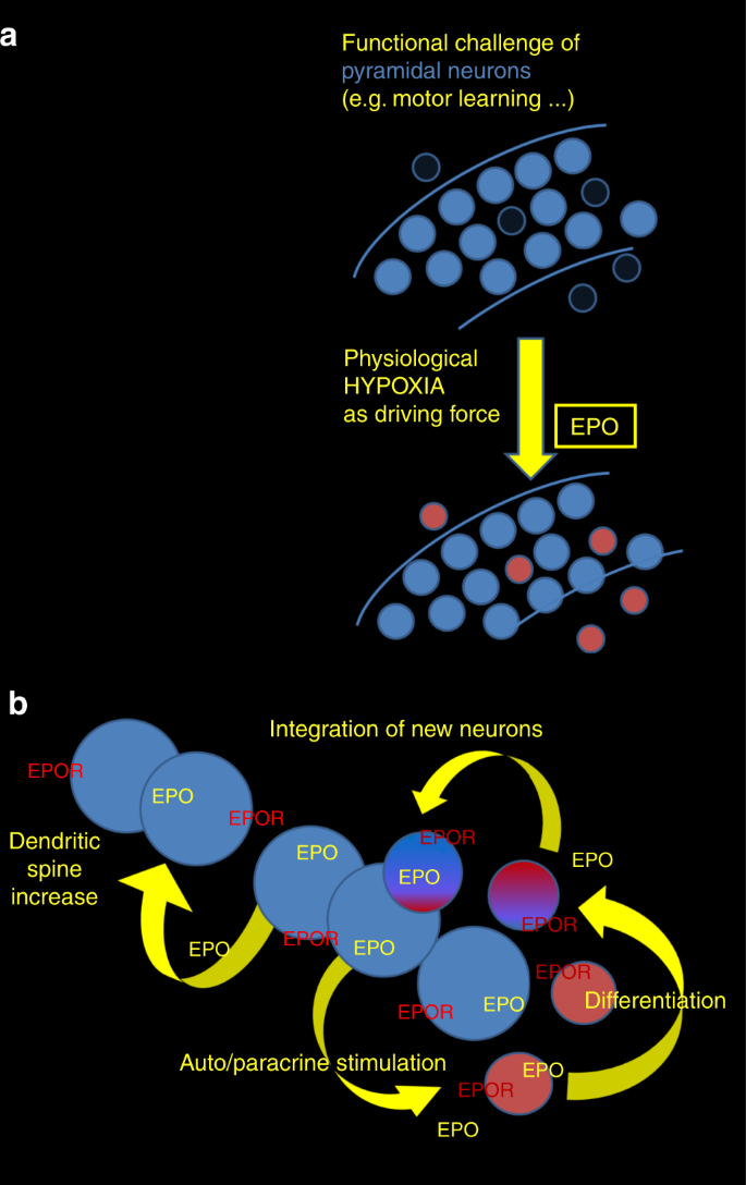
Taken together (Fig. 5), we have discovered that EPO – experimentally and clinically shown to improve cognition9,10,11,12,13,14,15,16,17 – stimulates a previously unrecognized mechanism of cellular neuroplasticity. Specifically, rhEPO (when peripherally administered) enters the brain, where it mimics the effects of endogenous neuronal EPO/EPOR signaling, stimulating dendritic spine formation on mature cells, and the differentiation of new pyramidal neurons from pre-existing (non-proliferating) precursors, the identity of which is currently unknown.
The EPO-responsive cells undergoing adult neurogenesis likely have no defining single genetic marker, suitable for CreER-based lineage tracing (unlike the EPO-responsive OPC17). Not even direct transdifferentiation of endothelial and astroglial cells or dedifferentiation of neurons can be completely excluded at this point. Jak2-Stat5 signaling, however, a pathway important for neuronal EPOR effects18,20, is known in various tissues to induce terminal differentiation, not dedifferentiation.
We hypothesize that the entire precursor cell lineage that is ready to differentiate toward pyramidal neurons in adult mice remains ‘in flow’. Similar to adaptive metabolic pathways, in which the ‘flux’ of metabolites matters more than the different steady-state concentrations, also in the proposed neuronal lineage progression, the EPO-responsive progenitor cells may never constitute abundant clusters in a cross-sectional steady-state analysis. This is remarkably similar to the effects of EPO on multiple precursors at different stages in the hematopoietic system1,30,31,32.
Pyramidal neurons, when challenged by novel tasks, undergo mild hypoxia, as detected here by ‘ODD labeling’, using hypoxia reporter mice. This activity-induced ‘functional hypoxia’ stimulates endogenous EPO expression by pyramidal neurons. In parallel, we find also EPOR expression enhanced, caused either directly by hypoxia33 or by EPO in an auto/paracrine manner34. When EPOR expression is deleted from pyramidal neurons, the endogenous EPO/EPOR system can no longer contribute to adaptive increase in performance.
Contrary to popular viewpoints that hypoxia is detrimental, recent reviews also consider beneficial effects and protection against cognitive dysfunction35,36. Our data using CRW indicate that hypoxia can act as driving force of long-lasting cellular neuroplasticity, complementing synaptic plasticity.
Neuronally produced EPO/EPOR likely constitutes an auto/paracrine mechanism, possibly in concert with other hypoxia-inducible genes, like vascular endothelial growth factor, which has previously also been reported to enhance cognition37. We suggest a model of an intrinsic EPO/EPOR-mediated mechanism of cellular neuroplasticity (Fig. 5), in form of a fundamental regulatory circle, which can explain the remarkable procognitive effects of rhEPO, consistently found in humans and rodents, health and disease9,10,12,14,15,16,38.
A recent study documented EPO mRNA in different neural cell types in the embryonic brain (mouse organogenesis cell atlas—MOCA39). The extraordinarily low expression of EPO and EPOR in the adult brain may explain why this system has previously not been rigorously studied. So far, we have focused on the hippocampal CA1 region, where we had obtained first evidence of substantially increased neuron numbers after rhEPO treatment17.
CA1 is involved in temporal pattern association and different facets of memory formation and consolidation40. Future studies will have to address EPO/EPOR functions in other CNS regions, including cortical areas known to be also engaged in CRW learning41, as well as neuron–glial interactions.
‘Adult neurogenesis’ from pre-existing precursors without proliferation extends the concept and pivotal work from many groups who discovered adult neurogenesis in distinct brain regions based on the selective labeling of proliferating cells in S-phase42,43,44,45,46,47,48. More complex than oligodendrogenesis from pre-existing precursors49, multiple neuronal progenitors may be relevant for maintaining neuron numbers in steady state, for adjustment to demand, and for rapid regenerative processes. The genetic approach of EPOR deletion in pyramidal neurons, together with the discovery of ‘functional hypoxia’ in the behaving brain, allowed us to propose a novel working model (Fig. 5). This model may add to our present concepts of neuroplasticity and adult neurogenesis.
reference link: https://www.nature.com/articles/s41467-020-15041-1
More information: Umer Javed Butt et al. Hippocampal neurons respond to brain activity with functional hypoxia, Molecular Psychiatry (2021). DOI: 10.1038/s41380-020-00988-w


