


 Extract (6-MSITC) in Healthy Older Adults")
: An In-Depth Exploration into its Thermogenic Role and Social Significance")


Last year, scientists at Scripps Research and Toscana Life Sciences studied the blood of 14 COVID-19 survivors to find the most potent antibodies against the SARS-CoV-2 virus.
One of the leading molecules that emerged – now in stage II/III trials in Italy – was an antibody dubbed J08, which seemed to be capable of both preventing and treating COVID-19.
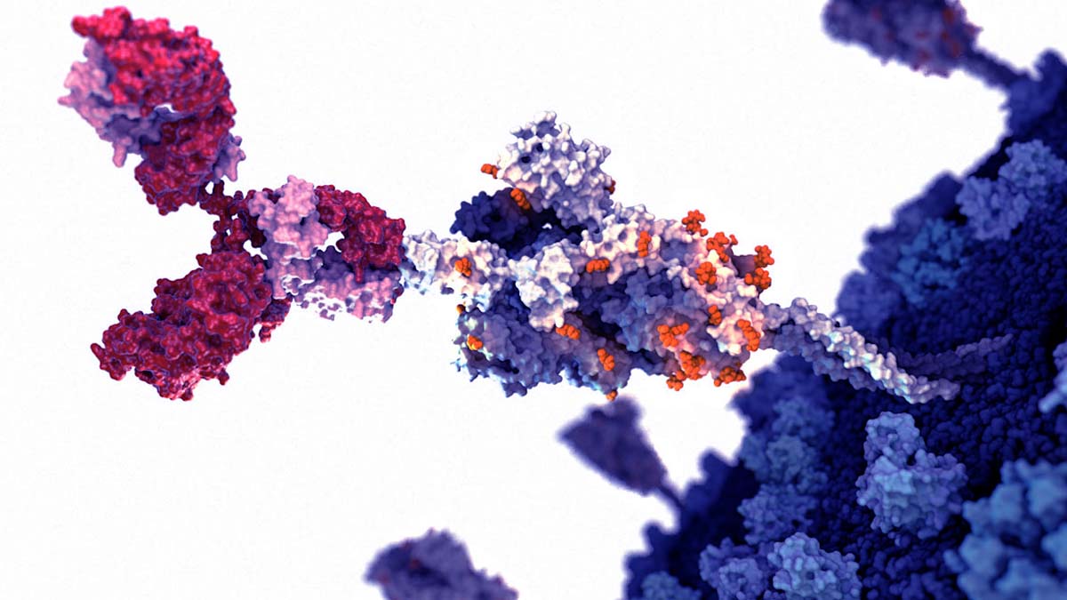
Now, the same group – a collaboration between scientists at Scripps Research and in Italy and France – has visualized exactly how J08 binds to different SARS-CoV-2 variants in different conformations, explaining what makes the monoclonal antibody so potent.
The research, published in Proceedings of the National Academy of Sciences, suggests that the J08 antibody, because of its flexibility, will likely remain effective against future variants of COVID-19.
When a person is exposed to a virus like SARS-CoV-2, their body generates a variety of antibodies that bind to different sections of the virus to clear it from the body. Scientists designing vaccines and treatments against COVID-19 are interested in what makes some of these naturally produced antibodies – like J08 – more effective than others.
In the months after Ward and his collaborators first identified J08, it became clear that the antibody, unlike many others, was potent against a variety of COVID-19 variants.
In the new work, the researchers determined the three-dimensional structure of J08 as it bound to the spike protein of SARS-CoV-2. They confirmed that J08 successfully attached to the Alpha, Beta, Gamma and Delta variants and neutralized the viruses—preventing them from replicating.
“With variants other than Omicron, this antibody binds quickly and doesn’t come off for hours and hours,” says co-first author Gabriel Ozorowski, a senior staff scientist in the Ward lab at Scripps Research. “With Omicron, we were initially happy to find that it still binds, but it falls off very quickly. We identified the two structural changes that cause this.”
The team showed that, for all the variants, J08 binds to a very small section of the virus – a section that generally stays the same even as the virus mutates.
“This small, flexible footprint is part of why J08 is able to withstand so many mutations—they don’t impact the antibody binding unless they happen to be in this one very small part of the virus,” says co-first author Jonathan Torres, lab manager of the Ward lab at Scripps Research.
The researchers say the new results support the continued clinical trials of the monoclonal antibody based on J08.
“I think we’re pretty confident that future variants won’t necessarily have both of these two critical mutations at the same time like Omicron,” says Ozorowski, “so that makes us hopeful that J08 will continue being very effective.”
SARS-CoV-2 neutralizing antibodies can be classified into four groups
Based on the first round of screening, 14 nAbs were selected for further characterization. All nAbs were able to bind the SARS-CoV-2 S protein in its trimeric conformation (Figure 3 A). The mAbs named J08, I14, F05, G12, C14, B07, I21, J13, and D14 were also able to specifically bind the S1 domain (Figure 3B). The nAbs named H20, I15, F10, and F20 were not able to bind single S1 or S2 domains but only the Sprotein in its trimeric state, while the nAb L19 bound only the S2 subunit (Figures 3B and 3C).
Among the group of S1-specific nAbs, only J08, I14, F05, G12, C14, and B07 were able to bind the S1 RBD and to strongly inhibit the interaction between the S protein and Vero E6 receptors, showing a half maximal effective concentration (EC50) at the NoB assay of 78.6, 15.6, and 68.5 ng/mL for J08-MUT, I14-MUT, and F05-MUT, respectively (Figures S3 A and S3B). On the other hand, I21, J13, and D14, despite showing S1 binding specificity, did not show any binding to the RBD and NoB activity (Figure S3A). Based on this description, four different groups of nAbs against SARS-CoV-2 were identified.
The first group (Group I) is composed of S1 RBD-specific nAbs (J08, I14, F05, G12, C14, and B07), which showed neutralization potency against the authentic wild type (WT), the D614G variant, and the emerging variant recently isolated in the UK B.1.1.7. S1 RBD-specific nAbs showing a neutralizing potency ranging from 3.9 to 157.5 ng/mL (Figures 3D–3I; Table S5) and picomolar affinity to the S protein with an equilibrium dissociation constant (KD) ranging from 0.2 to 4.6 E−10M (Figure S4 ). In addition to the D614G and the B.1.1.7 variants, the S1 RBD-specific nAb J08 showed also to neutralize SARS-CoV-2 variants containing the E484K mutation (Andreano et al., 2020). The second group (Group II) included S1-specific nAbs that did not bind the RBD (I21, J13, and D14).
These antibodies also showed good neutralization potency ranging from 99.2 to 500.0 ng/mL (Figures 3D–3I; Table S5) but inferior to that of S1 RBD-directed nAbs. One antibody from this group was not able to neutralize the B.1.1.7 variant (I21). The third group (Group III) is composed of antibodies able to bind the S-protein only in its whole trimeric conformation (H20, I15, F10, and F20). Antibodies belonging to this group showed lower affinity to the S protein trimer (KD 64.0 E−10M–757.0 E−10M) compared to Group I nAbs and medium neutralization potencies ranging from 155.0 to 492.2 ng/mL against the authentic WT and D614G (Figures 3D–3I; Figure S4; Table S5).
On the other hand, only one S protein-specific nAb (D21) showed moderate neutralization activity against the B.1.1.7 with an IC100 of 500.0 ng/mL. Three S protein-specific nAbs (I15, F10, and F20) did not show any functional activity against this latter variant (Figures 3D–3I; Table S5). The fourth and final group (Group IV) is composed of antibodies that exclusively recognized the S2 domain. Different antibodies with similar properties were identified for Group IV, but only the nAb L19 is shown. The Group IV nAb L19 shows the lowest neutralization potency with 19.8 μg/mL for the authentic WT, 12.5 μg/mL against the D614G, and 9.9 μg/mL against the B.1.1.7 variant (Figures 3D–3I; Table S5).

Functional characterization of potent SARS-CoV-2 S protein-specific nAbs
(A–C) Graphs show binding curves to the S protein in its trimeric conformation, S1 domain, and S2 domain. Mean ± SD of technical triplicates are shown. Dashed lines represent the threshold of positivity.
(D–F) Neutralization curves for selected antibodies were shown as percentage of viral neutralization against the authentic SARS-CoV-2 wild type (D), D614G variant (E), and the emerging variant B.1.1.7 (F). Data are representative of technical triplicates. A neutralizing COVID-19 convalescent plasma and an unrelated plasma were used as positive and negative control, respectively.
(G–I) Neutralization potency of 14 selected antibodies against the authentic SARS-CoV-2 wild type (G), D614G variant (H), and the emerging variant B.1.1.7 (I). Dashed lines show different ranges of neutralization potency (500, 100, and 10 ng/mL). In all graphs, selected antibodies are shown in dark red, pink, gray, and light blue based on their ability to recognize the SARS-CoV-2 S1 RBD, S1 domain, S protein trimer only, and S2 domain, respectively.

Binding to S protein receptor binding domain (RBD) and NoB activity of S1-RBD antibodies, related to Figure 3
(A) Histograms show the ability of selected antibodies to bind the S-protein RBD. Gray histograms represent the negative control while colored histograms show tested antibodies. Percentage of positive and negative populations are denoted on each graph.
(B) Neutralization of binding (NoB) curves for S1-RBD specific antibodies are shown as percentage of reduction of signal emitted by a fluorescently labled S-protein incubated with Vero E6 cells. Mean ± SD of technical duplicates are shown. Dashed lines represent the threshold of positivity; A neutralizing COVID-19 convalescent plasma and an unrelated plasma were used as positive and negative control, respectively.

Binding kinetics of SARS-CoV-2 nAbs to the S protein antigen, related to Figure 3
Representative binding curves of selected antibodies to SARS-CoV-2 S-protein trimer. Different curve colors define the spike concentration used in the experiment. Kon, Koff and KD are denoted on each graph.
All the antibodies described above were also tested for their ability to cross-neutralize other human coronavirus strains. nAbs were tested against lentiviral pseudotypes expressing the SARS-CoV-2, SARS-CoV-2 D614G, SARS-CoV, and Middle East respiratory syndrome (MERS)-CoV S protein on their viral membrane surface. Neutralization activity was shown against SARS-CoV-2 and D614G pseudotypes, therefore confirming previous data. None of the antibodies reported here were able to cross-neutralize other coronavirus species (Figure S5 ).

Neutralization activity of selected nAbs against SARS-CoV-2, SARS-CoV, and MERS-CoV pseudotypes, related to Figure 3
(A–D) Graphs show the neutralizing activities of 14 selected nAbs with different SARS-CoV-2 S-protein binding profiles against SARS-CoV-2, SARS-CoV-2 D614G, SARS-CoV and MERS-CoV pseudotypes respectively. Dashed lines represent the threshold of positivity. Mean ± SD of technical duplicates are shown. In all graphs selected antibodies are shown in dark red, pink, gray and light blue based on their ability to recognize the SARS-CoV-2 S1-RBD, S1-domain, S-protein trimer only and S2-domain respectively.
reference link : https://www.ncbi.nlm.nih.gov/pmc/articles/PMC7901298/
More information: Emanuele Andreano et al, Extremely potent human monoclonal antibodies from COVID-19 convalescent patients, Cell (2021). DOI: 10.1016/j.cell.2021.02.035
Jonathan L. Torres et al, Structural insights of a highly potent pan-neutralizing SARS-CoV-2 human monoclonal antibody, Proceedings of the National Academy of Sciences (2022). DOI: 10.1073/pnas.2120976119


