


 Extract (6-MSITC) in Healthy Older Adults")
: An In-Depth Exploration into its Thermogenic Role and Social Significance")


Doctors could soon get some help from an artificial intelligence tool when diagnosing brain aneurysms – bulges in blood vessels in the brain that can leak or burst open, potentially leading to stroke, brain damage or death.
The AI tool, developed by researchers at Stanford University and detailed in a paper published June 7 in JAMA Network Open, highlights areas of a brain scan that are likely to contain an aneurysm.
“There’s been a lot of concern about how machine learning will actually work within the medical field,” said Allison Park, a Stanford graduate student in statistics and co-lead author of the paper.
“This research is an example of how humans stay involved in the diagnostic process, aided by an artificial intelligence tool.”
This tool, which is built around an algorithm called HeadXNet, improved clinicians’ ability to correctly identify aneurysms at a level equivalent to finding six more aneurysms in 100 scans that contain aneurysms.
It also improved consensus among the interpreting clinicians.
While the success of HeadXNet in these experiments is promising, the team of researchers – who have expertise in machine learning, radiology and neurosurgery – cautions that further investigation is needed to evaluate generalizability of the AI tool prior to real-time clinical deployment given differences in scanner hardware and imaging protocols across different hospital centers.
The researchers plan to address such problems through multi-center collaboration.
Overview of the medical artificial intelligence (AI) research
Recently AI techniques have sent vast waves across healthcare, even fuelling an active discussion of whether AI doctors will eventually replace human physicians in the future. We believe that human physicians will not be replaced by machines in the foreseeable future, but AI can definitely assist physicians to make better clinical decisions or even replace human judgement in certain functional areas of healthcare (eg, radiology). The increasing availability of healthcare data and rapid development of big data analytic methods has made possible the recent successful applications of AI in healthcare. Guided by relevant clinical questions, powerful AI techniques can unlock clinically relevant information hidden in the massive amount of data, which in turn can assist clinical decision making.1–3
In this article, we survey the current status of AI in healthcare, as well as discuss its future. We first briefly review four relevant aspects from medical investigators’ perspectives:
- motivations of applying AI in healthcare
- data types that have be analysed by AI systems
- mechanisms that enable AI systems to generate clinical meaningful results
- disease types that the AI communities are currently tackling.
Motivation
The advantages of AI have been extensively discussed in the medical literature.3–5 AI can use sophisticated algorithms to ‘learn’ features from a large volume of healthcare data, and then use the obtained insights to assist clinical practice. It can also be equipped with learning and self-correcting abilities to improve its accuracy based on feedback. An AI system can assist physicians by providing up-to-date medical information from journals, textbooks and clinical practices to inform proper patient care.6 In addition, an AI system can help to reduce diagnostic and therapeutic errors that are inevitable in the human clinical practice.3 4 6–10 Moreover, an AI system extracts useful information from a large patient population to assist making real-time inferences for health risk alert and health outcome prediction.11
Healthcare data
Before AI systems can be deployed in healthcare applications, they need to be ‘trained’ through data that are generated from clinical activities, such as screening, diagnosis, treatment assignment and so on, so that they can learn similar groups of subjects, associations between subject features and outcomes of interest. These clinical data often exist in but not limited to the form of demographics, medical notes, electronic recordings from medical devices, physical examinations and clinical laboratory and images.12
Specifically, in the diagnosis stage, a substantial proportion of the AI literature analyses data from diagnosis imaging, genetic testing and electrodiagnosis (figure 1). For example, Jha and Topol urged radiologists to adopt AI technologies when analysing diagnostic images that contain vast data information.13 Li et al studied the uses of abnormal genetic expression in long non-coding RNAs to diagnose gastric cancer.14 Shin et al developed an electrodiagnosis support system for localising neural injury.15
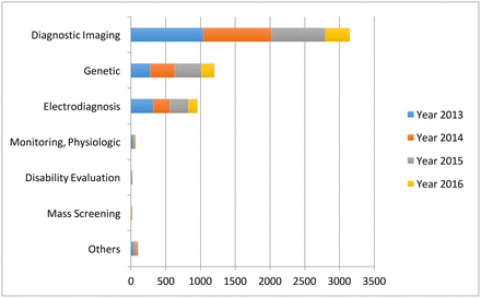
The data types considered in the artificial intelligence artificial (AI) literature. The comparison is obtained through searching the diagnosis techniques in the AI literature on the PubMed database.
In addition, physical examination notes and clinical laboratory results are the other two major data sources (figure 1).
We distinguish them with image, genetic and electrophysiological (EP) data because they contain large portions of unstructured narrative texts, such as clinical notes, that are not directly analysable.
As a consequence, the corresponding AI applications focus on first converting the unstructured text to machine-understandable electronic medical record (EMR).
For example, Karakülah et alused AI technologies to extract phenotypic features from case reports to enhance the diagnosis accuracy of the congenital anomalies.16
Augmented expertise
Combing brain scans for signs of an aneurysm can mean scrolling through hundreds of images.
Aneurysms come in many sizes and shapes and balloon out at tricky angles – some register as no more than a blip within the movie-like succession of images.
“Search for an aneurysm is one of the most labor-intensive and critical tasks radiologists undertake,” said Kristen Yeom, associate professor of radiology and co-senior author of the paper.
“Given inherent challenges of complex neurovascular anatomy and potential fatal outcome of a missed aneurysm, it prompted me to apply advances in computer science and vision to neuroimaging.”
Yeom brought the idea to the AI for Healthcare Bootcamp run by Stanford’s Machine Learning Group, which is led by Andrew Ng, adjunct professor of computer science and co-senior author of the paper.
The central challenge was creating an artificial intelligence tool that could accurately process these large stacks of 3-D images and complement clinical diagnostic practice.

To train their algorithm, Yeom worked with Park and Christopher Chute, a graduate student in computer science, and outlined clinically significant aneurysms detectable on 611 computerized tomography (CT) angiogram head scans.
“We labelled, by hand, every voxel – the 3-D equivalent to a pixel – with whether or not it was part of an aneurysm,” said Chute, who is also co-lead author of the paper.
“Building the training data was a pretty grueling task and there were a lot of data.”
Following the training, the algorithm decides for each voxel of a scan whether there is an aneurysm present.
The end result of the HeadXNet tool is the algorithm’s conclusions overlaid as a semi-transparent highlight on top of the scan.
This representation of the algorithm’s decision makes it easy for the clinicians to still see what the scans look like without HeadXNet’s input.
“We were interested how these scans with AI-added overlays would improve the performance of clinicians,” said Pranav Rajpurkar, a graduate student in computer science and co-lead author of the paper.
“Rather than just having the algorithm say that a scan contained an aneurysm, we were able to bring the exact locations of the aneurysms to the clinician’s attention.”
Eight clinicians tested HeadXNet by evaluating a set of 115 brain scans for aneurysm, once with the help of HeadXNet and once without. With the tool, the clinicians correctly identified more aneurysms, and therefore reduced the “miss” rate, and the clinicians were more likely to agree with one another.
HeadXNet did not influence how long it took the clinicians to decide on a diagnosis or their ability to correctly identify scans without aneurysms – a guard against telling someone they have an aneurysm when they don’t.
To other tasks and institutions
The machine learning methods at the heart of HeadXNet could likely be trained to identify other diseases inside and outside the brain.
For example, Yeom imagines a future version could focus on speeding up identifying aneurysms after they have burst, saving precious time in an urgent situation.
But a considerable hurdle remains in integrating any artificial intelligence medical tools with daily clinical workflow in radiology across hospitals.
Current scan viewers aren’t designed to work with deep learning assistance, so the researchers had to custom-build tools to integrate HeadXNet within scan viewers.
Similarly, variations in real-world data – as opposed to the data on which the algorithm is tested and trained – could reduce model performance.
If the algorithm processes data from different kinds of scanners or imaging protocols, or a patient population that wasn’t part of its original training, it might not work as expected.
“Because of these issues, I think deployment will come faster not with pure AI automation, but instead with AI and radiologists collaborating,” said Ng. “We still have technical and non-technical work to do, but we as a community will get there and AI-radiologist collaboration is the most promising path.”
More information: Allison Park et al. Deep Learning–Assisted Diagnosis of Cerebral Aneurysms Using the HeadXNet Model, JAMA Network Open(2019). DOI: 10.1001/jamanetworkopen.2019.5600
Journal information: JAMA Network Open
Provided by Stanford University


