


 Extract (6-MSITC) in Healthy Older Adults")
: An In-Depth Exploration into its Thermogenic Role and Social Significance")


Researchers have used artificial intelligence to support the instant diagnosis of one of the top causes of blindness, diabetes-related eye disease, in its earliest stages.
Diabetic retinopathy is the leading cause of vision loss in adults and its impact is growing worldwide, with 191 million people set to be affected by 2030.
There are no early-stage symptoms and the disease may already be advanced by the time people start losing their sight.
Early diagnosis and treatment can make a dramatic difference to how much vision a patient retains.
Now a team of Australian-Brazilian researchers led by RMIT University have developed an image-processing algorithm that can automatically detect one of the key signs of the disease, fluid on the retina, with an accuracy rate of 98%.
Lead investigator Professor Dinesh Kant Kumar, RMIT, said the method was instantaneous and cost-effective.
“We know that only half of those with diabetes have regular eye exams and one-third have never been checked,” Kumar said.
“But the gold standard methods of diagnosing diabetic retinopathy are invasive or expensive, and often unavailable in remote or developing parts of the world.
*-*-*-*-*-*
Overview
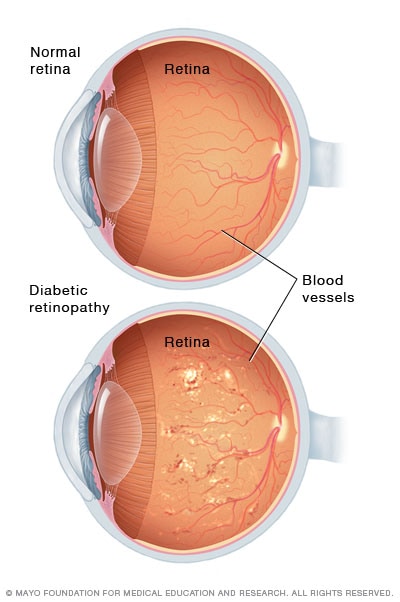
Diabetic retinopathy (die-uh-BET-ik ret-ih-NOP-uh-thee) is a diabetes complication that affects eyes. It’s caused by damage to the blood vessels of the light-sensitive tissue at the back of the eye (retina).
At first, diabetic retinopathy may cause no symptoms or only mild vision problems. Eventually, it can cause blindness.
The condition can develop in anyone who has type 1 or type 2 diabetes. The longer you have diabetes and the less controlled your blood sugar is, the more likely you are to develop this eye complication.
Symptoms
You might not have symptoms in the early stages of diabetic retinopathy. As the condition progresses, diabetic retinopathy symptoms may include:
- Spots or dark strings floating in your vision (floaters)
- Blurred vision
- Fluctuating vision
- Impaired color vision
- Dark or empty areas in your vision
- Vision loss
Diabetic retinopathy usually affects both eyes.
Causes
Over time, too much sugar in your blood can lead to the blockage of the tiny blood vessels that nourish the retina, cutting off its blood supply. As a result, the eye attempts to grow new blood vessels. But these new blood vessels don’t develop properly and can leak easily.
There are two types of diabetic retinopathy:
- Early diabetic retinopathy. In this more common form — called nonproliferative diabetic retinopathy (NPDR) — new blood vessels aren’t growing (proliferating).When you have NPDR, the walls of the blood vessels in your retina weaken. Tiny bulges (microaneurysms) protrude from the vessel walls of the smaller vessels, sometimes leaking fluid and blood into the retina. Larger retinal vessels can begin to dilate and become irregular in diameter, as well. NPDR can progress from mild to severe, as more blood vessels become blocked.Nerve fibers in the retina may begin to swell. Sometimes the central part of the retina (macula) begins to swell (macular edema), a condition that requires treatment.
- Advanced diabetic retinopathy. Diabetic retinopathy can progress to this more severe type, known as proliferative diabetic retinopathy. In this type, damaged blood vessels close off, causing the growth of new, abnormal blood vessels in the retina, and can leak into the clear, jelly-like substance that fills the center of your eye (vitreous).Eventually, scar tissue stimulated by the growth of new blood vessels may cause the retina to detach from the back of your eye. If the new blood vessels interfere with the normal flow of fluid out of the eye, pressure may build up in the eyeball. This can damage the nerve that carries images from your eye to your brain (optic nerve), resulting in glaucoma.
Risk factors
Anyone who has diabetes can develop diabetic retinopathy. Risk of developing the eye condition can increase as a result of:
- Duration of diabetes — the longer you have diabetes, the greater your risk of developing diabetic retinopathy
- Poor control of your blood sugar level
- High blood pressure
- High cholesterol
- Pregnancy
- Tobacco use
- Being African-American, Hispanic or Native American
Complications
Diabetic retinopathy involves the abnormal growth of blood vessels in the retina. Complications can lead to serious vision problems:
- Vitreous hemorrhage. The new blood vessels may bleed into the clear, jelly-like substance that fills the center of your eye. If the amount of bleeding is small, you might see only a few dark spots (floaters). In more-severe cases, blood can fill the vitreous cavity and completely block your vision.Vitreous hemorrhage by itself usually doesn’t cause permanent vision loss. The blood often clears from the eye within a few weeks or months. Unless your retina is damaged, your vision may return to its previous clarity.
- Retinal detachment. The abnormal blood vessels associated with diabetic retinopathy stimulate the growth of scar tissue, which can pull the retina away from the back of the eye. This may cause spots floating in your vision, flashes of light or severe vision loss.
- Glaucoma. New blood vessels may grow in the front part of your eye and interfere with the normal flow of fluid out of the eye, causing pressure in the eye to build up (glaucoma). This pressure can damage the nerve that carries images from your eye to your brain (optic nerve).
- Blindness. Eventually, diabetic retinopathy, glaucoma or both can lead to complete vision loss.
Prevention
You can’t always prevent diabetic retinopathy. However, regular eye exams, good control of your blood sugar and blood pressure, and early intervention for vision problems can help prevent severe vision loss.
If you have diabetes, reduce your risk of getting diabetic retinopathy by doing the following:
- Manage your diabetes. Make healthy eating and physical activity part of your daily routine. Try to get at least 150 minutes of moderate aerobic activity, such as walking, each week. Take oral diabetes medications or insulin as directed.
- Monitor your blood sugar level. You may need to check and record your blood sugar level several times a day — more-frequent measurements may be required if you’re ill or under stress. Ask your doctor how often you need to test your blood sugar.
- Ask your doctor about a glycosylated hemoglobin test. The glycosylated hemoglobin test, or hemoglobin A1C test, reflects your average blood sugar level for the two- to three-month period before the test. For most people, the A1C goal is to be under 7 percent.
- Keep your blood pressure and cholesterol under control. Eating healthy foods, exercising regularly and losing excess weight can help. Sometimes medication is needed, too.
- If you smoke or use other types of tobacco, ask your doctor to help you quit.Smoking increases your risk of various diabetes complications, including diabetic retinopathy.
- Pay attention to vision changes. Contact your eye doctor right away if you experience sudden vision changes or your vision becomes blurry, spotty or hazy.
Remember, diabetes doesn’t necessarily lead to vision loss. Taking an active role in diabetes management can go a long way toward preventing complications.
*-*-*-*-*-*
“Our AI-driven approach delivers results that are just as accurate as clinical scans but relies on retinal images that can be generated with ordinary optometry equipment.
“Making it quicker and cheaper to detect this incurable disease could be life changing for the millions of people who are currently undiagnosed and risk losing their sight.”

Fluorescein angiography and optical coherence tomography scans are currently the most accurate clinical methods for diagnosing diabetic retinopathy.
An alternative and cheaper method is analysing images of the retina that can be taken with relatively inexpensive equipment called fundus cameras, but the process is manual, time-consuming and less reliable.
To automate the analysis of fundus images, researchers in the Biosignals Laboratory in the School of Engineering at RMIT, together with collaborators in Brazil, used deep learning and artificial intelligence techniques.
The algorithm they developed can accurately and reliably spot the presence of fluid from damaged blood vessels, or exudate, inside the retina.
The researchers hope their method could eventually be used for widespread screening of at-risk populations.
“Undiagnosed diabetes is a massive health problem here and around the globe,” Kumar said.
“For every single person in Australia who knows they have diabetes, another is living with diabetes but isn’t diagnosed. In developing countries, the ratio is one diagnosed to four undiagnosed.
“This results in millions of people developing preventable and treatable complications from diabetes-related diseases.
“With further development, our technology has the potential to reduce that burden.”
The researchers are in discussions with manufacturers of fundus cameras about potential collaborations to advance the technology.
More information: Parham Khojasteh et al, Exudate detection in fundus images using deeply-learnable features, Computers in Biology and Medicine (2018). DOI: 10.1016/j.compbiomed.2018.10.031
Provided by RMIT University


