


 Extract (6-MSITC) in Healthy Older Adults")
: An In-Depth Exploration into its Thermogenic Role and Social Significance")


Researchers led by biologists at Tufts University have discovered that the brains of developing embryos provide signals to a nascent immune system that help it ward off infections and significantly improve the embryo’s ability to survive a bacterial challenge.
Using frog embryos, which continue to develop with their brains removed, the researchers found that embryos without a brain are not able to marshall the forces of immune cells to an injury or infection site, leading the embryo to succumb to an infection more quickly.
By contrast, the presence of a brain crucially helps direct immune cells to the site of injury to overcome the bacterial threat.
The study was published today in NPJ Regenerative Medicine.
In a developing embryo, both brain and immune system are not fully formed.
The immune system, for its part, consists mostly of an “innate” system of cells that respond immediately to infection and do not require training or produce antibodies.
Nevertheless, these cells require signals that prompt them to move toward an infection site and trigger a response.
The research team found that the brain appears to contribute to the signals that guide the nascent immune system.
When brainless frog embryos were infected with E. coli, only about 16% of embryos survived, while the presence of a brain protected more than 50% from the infection.
By following markers of immune cells, researchers confirmed that the effect is not due to the missing brain somehow hampering immune system development because the composition of the immune cells remained the same with or without a brain.
Instead, they found that the effect was due to the brain sending signals to the immune cells to move toward the site of an infection.
“We found that macrophages –innate immune system cells that can swallow up bacteria and destroy them to reduce the burden of an infection do not migrate appropriately without the presence of the brain” said Michael Levin, Vannevar Bush Professor of Biology at Tufts University’s School or Arts and Sciences and Associate Faculty at Harvard’s Wyss Institute, director of the Allen Discovery Center at Tufts and corresponding author of the study. “Without the brain and its neurotransmitter signals, gene expression and innate immune system activity go awry, resulting in increased susceptibility to bacterial pathogens. ”
Other roles for the embryonic brain signaling during infection may include inducing cellular responses, for example preventing cell death or reducing inflammation, that help protect against the harmful effects of the infection.
Tail of a Xenopus laevis (frog) embryo developed without brain showing an aberrant distribution of immune cells (macrophages in green) lying in close proximity to the chaotically-sprouted peripheral neural network (nerves in red).
Immune system abberations were also observed in brainless embryos that were further developed.
When the researchers tracked myeloid cells, a class of immune cells that includes macrophages, neutrophils and others, after an injury, they found that the myeloid cells in brainless embryos gathered in locations far from the injury site.
By contrast, myeloid cells in normal embryos with intact brains would pile up at the injury site to assist in healing.
In fact, in the brainless embryo, the myeloid cells tended to cluster around abnormal, disorganized peripheral nerve networks, also a by-product of brain absence, as demonstrated in earlier studies.
An examination of aberrations in genetic expression in brainless embryos also pointed to the reduction of the neurotransmitter dopamine (a signaling chemical used in the brain for learning and motivation), and that dopamine may play a role in activating immune cells to migrate in the early stages of an infection.
The absence of an immune cell quorum at the infection site leads the brainless embryos to become more susectible to its lethal effects.

Face of a Xenopus laevis (frog) embryo developed with brain, showing parts of the central nervous system in green (brain on the top down to the middle and retina of the eye to the left) and cranial nerves in red. Image is credited to Michael Levin.
“Our results demonstrate the deep interconnections within the bacteria-brain-body axis: the early brain is able to ‘sense’ the pathogenic bacteria and to elaborate a response targeted to fight against the cellular and molecular consequences of the infection,” said Celia Herrera Rincon, Research Scientist II at the Allen Discovery Center at Tufts, and first author of the study.
Other authors of this study include: Jean-Francois Paré, Christina Harrison, Alina Fischer, and Sophia Jannetty at the Allen Discovery Center at Tufts; Christopher Martyniuk, associate professor in the Department of Physiological Sciences at University of Florida; and Alexandre Dinis and Vishal Keshari, graduate students, and Richard Novak, senior staff engineer at the Wyss Institute for Biologically Inspired Engineering, Harvard Universiy.
Funding: This research was supported by the Templeton World Charity Foundation Independent Research Fellowship (TWCF0241) and the Allen Discovery Center program through The Paul G. Allen Frontiers Group (12171), as well as The Defense Advanced Research Projects Agency (DARPA, W911NF-16-C-0050), and the National Institutes of Health (AR055993, AR061988). The content is solely the responsibility of the authors and does not necessarily represent the official views of the National Institutes of Health.
Herrera-Rincon, C., Paré, J-F, Martyniuk, C.J., Jannetty, S.K., Harrison, C., Fischer, A., Dinis, A., Keshari, V., Novak, R., and Levin, M. “An in vivo brain–bacteria interface:
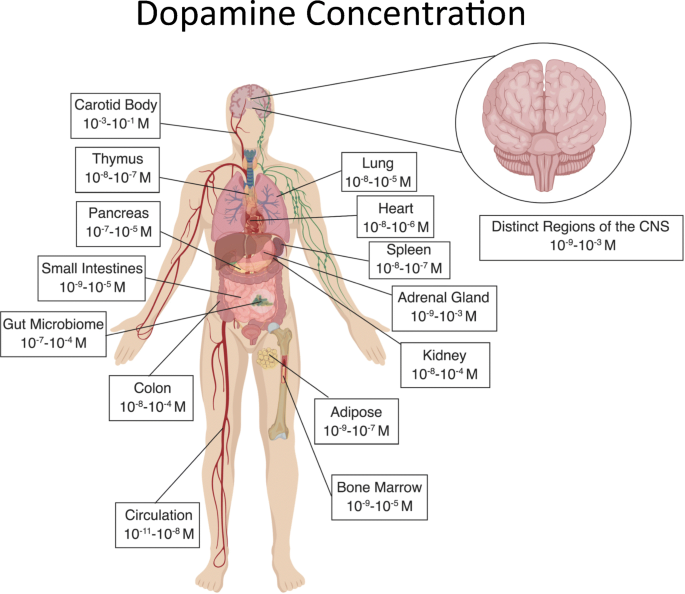
Dopamine is well recognized as a neurotransmitter in the brain, and regulates critical functions in a variety of peripheral systems.
Growing research has also shown that dopamine acts as an important regulator of immune function. Many immune cells express dopamine receptors and other dopamine related proteins, enabling them to actively respond to dopamine and suggesting that dopaminergic immunoregulation is an important part of proper immune function.
A detailed understanding of the physiological concentrations of dopamine in specific regions of the human body, particularly in peripheral systems, is critical to the development of hypotheses and experiments examining the effects of physiologically relevant dopamine concentrations on immune cells.
Unfortunately, the dopamine concentrations to which these immune cells would be exposed in different anatomical regions are not clear.
To address this issue, this comprehensive review details the current information regarding concentrations of dopamine found in both the central nervous system and in many regions of the periphery.
In addition, we discuss the immune cells present in each region, and how these could interact with dopamine in each compartment described.
Finally, the review briefly addresses how changes in these dopamine concentrations could influence immune cell dysfunction in several disease states including Parkinson’s disease, multiple sclerosis, rheumatoid arthritis, inflammatory bowel disease, as well as the collection of pathologies, cognitive and motor symptoms associated with HIV infection in the central nervous system, known as NeuroHIV.
These data will improve our understanding of the interactions between the dopaminergic and immune systems during both homeostatic function and in disease, clarify the effects of existing dopaminergic drugs and promote the creation of new therapeutic strategies based on manipulating immune function through dopaminergic signaling.
Source:
Tufts


