


 Extract (6-MSITC) in Healthy Older Adults")
: An In-Depth Exploration into its Thermogenic Role and Social Significance")


Bones grow curved in response to pressure and strain and these changes can be long-lasting, according to a new study by UCL, the Royal Veterinary College London and the Max Planck Institute.
Bones can change their shape throughout our life by regulating bone formation and resorption processes, which is often a response to forces which press, pull and twist the skeleton during everyday movements and exercise. The purpose of this shaping is to limit any risk of fracture.
Understanding that a bone’s fracture resistance is based on engineering rules which would predict a completely straight shape to be optimal, the research team sought to understand why most of our bones are curved if the goal of these changes in shape is to prevent fracture.
Whilst undertaking this study, the team knew that other important questions would likely also be answered, for instance, learning whether the beneficial effects of exercise on our skeletons might be long-lived.
Researchers from the RVC used 4-D in vivo high-resolution micro-computed tomography and computational methods to monitor shape changes in an entire bone over an extended period after highly controlled exposure to a known force.
The team also developed a novel approach to quantify 3-D bone formation and resorption surfaces validated by conventional histology and how these relate to local stress in bone tissue caused by physiological force.
The research, published in Science Advances, was undertaken to reveal the location and extent of force-related resorption and formation at the whole bone level and to determine if the changes are, as has been predicted for many years, rapidly reversible.
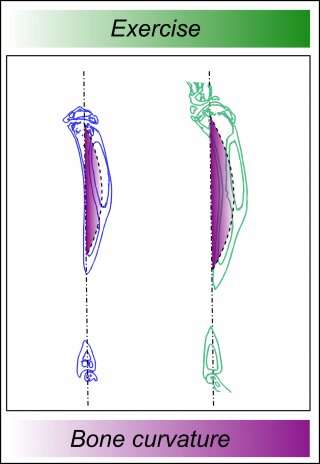
The results indicate that the bone’s response to these forces varies along its length to make the bone more curved in most parts and that these shape changes are very long-lived.
The researchers found that the curving process needed to be highly targeted; some short-term gains were rapidly lost through resorption, but others were preserved.
From precise mapping of the change over-time, researchers showed that the reversible and preserved bone changes were distinguishable on the basis that they initially targeted either local resorption or formation.
It was a surprise to the team that the preserved increases in bone curvature operate independently of local stress levels.
The findings provide experimental evidence by which “Harold Frost’s long-held theoretical framework’ can be adjusted with an added hierarchy based upon fracture resistance. While some benefits of exercise-related load will gradually disappear, others will be preferentially retained.
They also indicate that increased curvature engenders a built-in warning mechanism predicting how best to respond to physiological forces in the future.
The changes in curvature do not compromise the strength, as increased quantity compensates for this beneficial change in bone shape.
Professor Peter Lee (UCL Mechanical Engineering) said: “Multi-disciplinary teams at the RVC, UCL and other multi-disciplinary groups have been collaborating for many years to use high resolution laboratory and synchrotron X-ray tomography to try and understand how skeletal disorders and bone architecture interact.
This most recent study demonstrates the importance of in situ loading studies in helping us to understand how bones bear load both in terms of health, skeletal disease progression and fracture risk.”
Dr. Behzad Javaheri, Postdoctoral Research Associate at the RVC, said: “This study implies that new ‘intelligent’ bone therapies, which interact with physiological forces to selectively preserve overall curvature to resist fracture, would be more desirable.
Existing osteoporosis treatments do not currently target the bone regions that most dramatically contribute to bending strength; they instead have generalised anti-resorptive/pro-formative effects.
“The research emphasises that osteoporosis trials may be improved by respecting the mechanical origin of the enhanced fracture risk, rather than simply focussing on commonly affected sites; failure to retain appropriate curvature levels may dictate osteoporotic fracture risk.
“Follow-up tracking of bone shape modifications after orthopaedic surgery may also be beneficial and a more long-term view should be taken to monitoring the effects of exercise on skeletal health.”
Dr. Hajar Razi, Postdoctoral Researcher at the Max Planck Institute of Colloids and Interfaces, said: “We have known for quite some time that bone adapts to mechanical forces.
Here in this study we show that, at the organ-level, this adaptation is non-linear with respect to the local mechanical stimuli.
In fact, bone response to mechanical stimulation we observed implies an adaptation to a bigger goal: to achieve a larger bone curvature adjusted for load predictability.”
A fundamental tenet of bone biomechanics is the adaptation phenomenon of bone microstructure under regularly applied mechanical loading1,2. It has been hypothesized by “Wolff’s law”3 that this dynamic adaptive process, termed bone remodeling, occurs in bone mass and architecture due to stimuli obtained from its mechanical environment4,5.
Bones can receive stimuli in the form of mechanical loading resulting from various intensities in physical activities and sports. Ground reaction forces, which generate stresses and strains on our weight-bearing bones, are determinant factors of bone remodeling.
They are greater when we move faster and/or more intensively, hence vary according to the type of physical activities6. Low impact sports such as swimming, can produce lower ground reaction forces than high impact sports such as gymnastics or cross-country running7,8.
A regular and sufficient amount of impact loading can prove to be very effective for bones at all ages9.
Well-controlled and physiologic mechanical loading models are essential to successfully define and identify the anabolic and catabolic mechanisms involved in bone remodeling. To date, many loading models have been implemented in different animal studies, ranging from whole body rodent vibration, exercise models, and in vivo bone loading models such as tibial bending, ulnar and tibial axial loading10–14.
Compared to other models, the tibial compression model has the potential to generate cortical and trabecular bone adaptation under applied mechanical load15,16. Axial loading in mouse tibia has been used for several years to investigate the effects of loading as a function of age17,18, sex19, disease20 and strain level16.
However, still, there is a lack of data on the loading mechanism and effects of non-invasive loading in bone formation for the rat tibial axial compression model.
Adolescence is a dynamic period for bone growth and development21. In this period, regular impact loading in sufficient amount can ensure a proper bone accrual and also contribute to building up a strong skeleton22.
It has been reported that23 performing a high impact physical activity, such as jumping, can effectively contribute to improving the hip bone strength in adolescents24. There are a few clinical and animal studies investigating the effects of impact loading on adolescent growth, but results are inconsistent.
In some published clinical studies25,26, researchers have not provided a clear, distinct separation between the nutritional and mechanical factors. The studies expressed the physical activities in hours per week whereas the intensity of the activity (repetitions, peak load, and frequency) have not been considered.
As a result, it becomes challenging to infer about the isolated effect of impact loading on the growing bones from these studies. In animal studies, the effects of exercise on long bone growth are also inconsistent, resulting in either no change27, minimal change28, or significant reduction29,30 in growth, measured as changes in long bone length.
Hence, if any loading induced changes occur during the adolescence and whether they modify the bone microstructure are clinically relevant questions which remain still unresolved. We hypothesize that bone morphometry and biomechanics can be improved, but bone growth would remain unaffected under the controlled impact loading applied during the adolescence period.
Moreover, among three different impact loading levels, we suggest that the higher impact intensity influences more importantly bone morphometry and biomechanics. This study aimed to use an animal model (rat tibia) to investigate the effects of well controlled in vivo low, medium and high impact loadings applied during puberty on bone growth, morphometry and biomechanics.
More information: Behzad Javaheri et al. Lasting organ-level bone mechanoadaptation is unrelated to local strain, Science Advances (2020). DOI: 10.1126/sciadv.aax8301


