


 Extract (6-MSITC) in Healthy Older Adults")
: An In-Depth Exploration into its Thermogenic Role and Social Significance")


Despite significant advances in prevention and treatment of cancer, year after year there is a steady increase in the number of deaths caused by malignancy.
In industrialised nations, one in every two or three people develops a form of cancer during the course of their life, and the trend is rising.
For a long time, certain pathogens, in particular viruses and bacteria, have been considered as potential causes of cancer.
Three viruses and one bacterium are recognized as risk factors: the human papillomavirus, the human hepatitis viruses B and C, as well as the bacterium Helicobacter pylori usually found in the stomach.
But in addition to these well-known pathogens, does a healthy microbiome, i.e. the entirety and balance of microorganisms in the body, also play a role in the development of cancer?
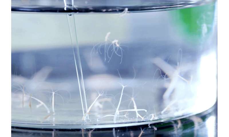
A research team led by Professor Thomas Bosch at Kiel University has demonstrated, using the simple freshwater polyp Hydra as an experimental model, that after an environmental disruption of the normal bacterial community, the tissue of a host organism may be colonized by bacteria from the environment.
The contact with already-present microbes then leads to the production of bacterial factors that have a damaging effect on the Hydra cell structures, and ultimately trigger tumor formation. The Kiel team published their research results, which were achieved in frame of the Collaborative Research Centre (CRC) 1182 “Origin and Function of Metaorganisms” at Kiel University, today in the renowned scientific journal PLOS Pathogens.
Evolutionary origins of cancer
The research team was prompted to use the evolutionary approach to cancer by their previous findings from work on the origins of cancer during the evolutionary history of life. Several years ago, the researchers already used the freshwater polyp Hydra, a phylogenetically ancient multicellular organism, to demonstrate that in principle all multicellular animals can form tumors.
“We believe that cancer is a legacy of transition to multicellularity early in the evolution of life,” emphasised Dr. Alexander Klimovich, a researcher in the field of cell and developmental biology at Kiel University and leader of the study.
“Since all multicellular organisms also possess a microbiome, and have evolved together with their microbial symbionts over millions of years, it is logical to hypothesize that the microorganisms are also involved in the development of cancer,” continued Klimovich.
Disrupted bacterial colonization triggers tumor formation
In laboratory experiments, the doctoral researcher Kai Rathje, a member of the research team, has now been able demonstrate such a causal involvement of individual bacterial species and their interactions within the microbiome in cnidarians.
“Hydra develop cancer if a specific type of foreign bacteria from the phylum Spirochaetes becomes increasingly prevalent in the microbiome, and thereby disrupts the balance of the bacterial colonization in their tissue,” emphasized Klimovich.
“Interestingly, these bacteria exert their harmful influence only in the presence of certain other bacteria from the genus Pseudomonas, which are part of the normal composition of the microbiome,” Klimovich continued.
Thus, the interaction of microbes with each other is involved in tumor formation in cnidarians, and the initial impulse also comes from the influence of the environment: the organisms initially acquire the harmful spirochaetes from the surrounding water. However, the invading bacteria successfully colonize the host tissue only if the Hydra tissue has already been weakened by changing factors in the environment.
These factors include a change in temperature, and the resulting change in microbial colonization. The researchers were able to prove experimentally that spirochaetes and Pseudomonas bacteria from the natural microbiome interact with each other, and thereby both change their behavior dramatically. When they encounter each other, the bacteria modify their movement patterns and seek direct contact with one another.
As a result, they also begin to express different genetic information and thereby in particular activate factors which have a pathogenic effect for the host organism. Due to these changes, the microbial balance in the tissue of the cnidarians becomes disrupted, which is followed by structural changes in the cells and ultimately to tumor formation.
How these interactions occur at the molecular level, and which specific biochemical mechanisms are involved in this form of cancer development, are the subject of currently ongoing investigations.
The microbiome-gateway and protective barrier at the same time
“Our novel findings point to a universal principle which will expand our understanding of the pathogenesis of cancer-namely, as a comprehensive interaction of genetic and environmental factors, including specific microbial influences,” emphasized Professor Thomas Bosch, head of cell and developmental biology at Kiel University and spokesperson for the CRC 1182.
“The new research findings show that an important aspect in the pathogenesis of cancer can be found in the context of microbial interactions – it was only the common presence of certain bacteria interacting with each other within a disturbed microbiome that enabled the formation of tumors in the case we investigated. Therefore, in many cases, it is probably not a single malicious intruder, but the malfunction of the microbiome as a protective barrier for the body as a whole what can promote the development of cancer,” added Bosch.
These findings offer a promising perspective because the protective function of the microbiome could possibly be used in future: “The microbial colonization of the body normally balances out and protects the organism against harmful influences, potentially even against carcinogenic influences,” said Bosch.
“Future research will show whether this ability of the microbiome to maintain a healthy barrier which protects the body from colonization by harmful microorganisms may also be used for the prevention of cancer,” continued Bosch.
In the future, there could be targeted interventions in the composition of the microbiome. Such manipulations could potentially hinder the establishment of certain cancer-promoting bacterial communities, and thus restore the healthy balance of the microbiome. However, more extensive fundamental research is required before such preventive measures or treatments can be implemented.
Evidence linking the gut microbiome to cancer immunotherapy
Multiple studies support that gut microbes can profoundly influence the potency of immunotherapy and some chemotherapies with immunostimulatory functions (summarized in Table 1).
Pioneering work in this field found that intestinal microbiota was essential for optimal responses to CpG-oligonucleotide immunotherapy which activates innate immune cells through TLR9 [85].
Similarly, the gut microbiota was found to shape the anti-cancer immune response by stimulating generation of a specific subset of “pathogenic” Th17 (pTh17) cells and memory Th1 immune response after treatment with immune-stimulatory chemotherapy cyclophosphamide [86].
Certain bacterial taxa in patients with hematologic malignancies are associated with efficacy of allogeneic hematopoietic stem cell transplantation (allo-HSCT) and decreased risk for graft-versus-host disease (GVHD) following therapy [87, 88].
Initial evidence for the contribution of specific microbes to immune checkpoint blockade (ICB) immunotherapy, including CTLA-4 and PD-1/PD-L1 blockade, was demonstrated in mouse models [17, 18]. B. fragilis was reported to enhance anti-CTLA-4 efficacy via a proposed mechanism involving the activation of Th1 cells with cross-reactivity to bacterial antigens and tumor neoantigens [18].
Oral administration of Bifidobacterium increased tumor infiltration and IFN-γ production by CD8+ tumor-specific T cells and improved both basal tumor control and anti-PD-L1 efficacy via a proposed mechanism involving increased activation of splenic and intratumoral DCs [17].
These mouse studies established the importance of the microbiome in cancer ICB therapy and inspired clinical pursuits to assess the microbiome’s impact on anti-CTLA-4 and anti-PD-1/PD-L1-based therapies in patients.
Table 1
Studies linking the gut microbiome composition to efficacy of cancer therapy. The table summarizes major findings from clinical and preclinical studies pointing to a link between gut bacteria and therapeutic outcomes in the context of various cancers and therapeutic regimens
| Major finding | Mouse or Human data | Cancer/Therapy | Reference |
|---|---|---|---|
| Chemotherapy with immunostimulatory properties | |||
| Akkermansia muciniphila abundance in baseline stool samples was associated with response to ICB | Mouse | Various cancer models/Cyclophosphamide immunostimulatory chemotherapy | [86] |
| Presence of intratumoral gammaproteobacteria was associated with resistance to gemcitabine chemotherapy | Human; Mouse | Pancreatic ductal adenocarcinoma/ Gemcitabine immunostimulatory chemotherapy | [94] |
| Immunotherapy | |||
| Commensal microbiota was required for optimal response to therapy | Mouse | Various cancer models/ CpG-oligonucleotide + anti-IL-10R antibody and platinum chemotherapy (oxaliplatin) | [85] |
| Total body irradiation disrupted intestinal barrier and improved outcome of T-cell based therapy by a mechanism dependent on LPS/microbe translocation and TLR4 signaling | Mouse | Melanoma/Adoptive T cell transfer | [97] |
| Eubacterium limosum abundance was associated with decreased risk of relapse or disease progression | Human | Hematologic cancers/Allo-HSCT | [88] |
| Blautia abundance was associated with increased overall survival and reduced risk of GVHD | Human | Hematologic cancers/Allo-HSCT | [87] |
| Bacteroides abundance was associated with resistance to ICB-induced colitis | Human | Metastatic melanoma/Anti-CTLA-4 | [93] |
| Bacteroides abundance was associated with response to ICB | Mouse; Human | Metastatic melanoma/Anti-CTLA-4 | [18] |
| Bifidobacterium abundance was associated with improved spontaneous anti-tumor immunity and response to ICB | Mouse | Melanoma/Anti-PD-L1 | [17] |
| Faecalibacterium and other Firmicutes abundance in baseline stool samples was associated with response to ICB; Bacteroides abundance was associated with poor responsiveness to ICB | Human | Metastatic melanoma/Anti-CTLA-4 | [92] |
| Bacteroides caccae, Faecalibacterium prausnitzii, Bacteroides thetaiotaomicron, Holdemania filiformis, and Dorea formicogenerans were associated with response to ICB | Human | Metastatic melanoma/Anti-PD-1; Anti-CTLA-4 | [91] |
| A. muciniphila abundance in baseline stool samples was associated with response to ICB | Human; Mouse | Non-small cell lung cancer; Renal cell carcinoma/Anti-PD-1 | [89] |
| Higher microbiome richness, Clostridiales, Ruminococcaceae, and Faecalibacterium abundance, and enrichment in genes involved in anabolic pathways in baseline stool samples were associated with responsiveness to ICB | Human; Mouse | Metastatic melanoma/Anti-PD-1 | [90] |
| Several dozen bacterial species in baseline stool samples were differentially enriched between patients with strong vs. poor responsiveness to ICB | Human; Mouse | Metastatic melanoma/Anti-PD-1 | [44] |
Results from multiple institutions have contributed to the growing consensus that the gut microbiome is linked to immunotherapy efficacy in cancer patients [44, 89–92]. DNA sequencing of stool samples collected prior to checkpoint blockade therapy identified an association between gut microbiome composition and subsequent therapeutic response.
Distinct bacterial taxa were overrepresented in responder (R) patients, whereas other bacterial sequences were over-represented in non-responder (NR) patients. Importantly, only some of these identified bacteria were consistent across multiple studies.
This discrepancy may reflect discordant biology-the patient populations were from geographically distinct locations, with potentially dissimilar environmental and genetic factors-but also may be explained by technical differences, such as fecal collection, storage and DNA extraction and sequencing methods, as well as downstream bioinformatic analysis.
Moving beyond correlative studies, human microbiota “avatars” (GF mice colonized with patient stool-derived commensals) have been used to show the mechanistic contribution of the microbiota to treatment response.
Mirroring patient data, mice reconstituted with R patient fecal material showed greater benefit from checkpoint blockade than mice colonized with NR fecal samples [44, 89, 90].
Beyond clinical efficacy rate, immune-related toxicity of ICB has also been linked to the composition of the gut microbiome. Based on stool samples collected from patients treated with an anti-CTLA-4 antibody, bacteria in the Bacteroidetes phylum were associated with lower incidence of treatment-induced colitis [93].
Deciphering the biological mechanism of microbiome mediated immune modulation
These findings linking the gut microbiome to immunotherapy efficacy only scratch the surface of this complex interaction. Determining the biological mechanisms is critical for moving towards therapeutic manipulation of the microbiota to optimize patient response. Tractable mouse models are being utilized to explore the causal role gut bacteria play in treatment efficacy.
When it comes to exploring the possible mechanisms of microbiota-mediated modulation of anti-tumor immunity, two general questions arise.
First, what is the nature of the messenger, which delivers a signal from the GI tract to the tumor and/or tumor-draining lymph node (TdLN)?
Such a messenger would be able to enter the circulation in order to access the distant tumor site and can be classified as microbiota- or host-derived cell (live microbes or host immune cells) or molecule (MAMP/PAMP, microbial metabolite, or host cytokine).
The second question is what is the nature of the immune effect that the messenger confers within the tumor?
An immunosuppressive effect could be achieved by augmenting regulatory functions (Tregs, MDSCs, tumor-associated macrophages) or directly inhibiting anti-tumor immunity; an immunostimulatory effect could be achieved by alleviating regulatory functions or stimulating anti-tumor T cell responses (via antigenicity, adjuvanticity or bystander activation).
The exact mechanisms of microbiota-mediated effects on tumor growth and efficacy of immunotherapy are only beginning to be understood. Figure 1 summarizes these hypothetical scenarios and early evidence is discussed below.

Possible mechanisms linking the gut microbiota to anti-tumor immunity. The composition of the gut microbiome may impact immunotherapy efficacy by either acting as (1) an immunosuppressive or (2) an immunostimulatory factor via various non-mutually exclusive mechanisms. (1) Certain commensal bacteria may suppress anti-tumor immunity by skewing immune subset balances towards suppressive phenotypes such as Tregs and MDSCs. Locally in mucosal sites, induction of immunosuppressive cells could be mediated by cytokines released by host cells (such as gut epithelium or immune cells) in response to microbial sensing. Immunosuppressive effects in distant sites, such as active immunosuppression in the TME, could be mediated by trafficking of locally induced suppressor cells. Additionally, bacterial metabolites with immunosuppressive properties might be released into the circulation and promote immunosuppressive cell functions in the TdLN and TME. Chronic inflammation caused by continuous stimulation by PAMPs/MAMPs or epithelial injury could also ultimately contribute to immunosuppression over time. (2) The immunostimulatory effects of the gut microbiota could be mediated by augmented antigenicity, adjuvanticity, or bystander T cell activation. (a) Antigenicity: Cross-reactive T cells driven by bacterial antigens that additionally recognize tumor-associated antigens is one conceivable mechanism. Luminal bacteria or bacterial antigens can be internalized by DCs in the LP via trans-endothelial dendrites extending through the epithelium into the lumen. Goblet cells and M cells can also serve as portals to deliver bacterial antigens to mucosal APCs. Alternatively, disruption of barrier function may allow for the translocation of luminal bacteria and bacterial antigens. Antigen-loaded DCs can migrate from the LP to the MLN and possibly to distant sites such as the TdLN, where they may prime cross-reactive anti-tumor CD8+ or CD4+ T cells, enhancing cytotoxic T lymphocyte (CTL) function in the TME. (b) Adjuvanticity: PAMPs/MAMPs may condition DCs to be more potent T cell activators, for instance by upregulating costimulatory molecule expression, enhancing antigen presentation, or boosting type I IFN production. Some microbial metabolites could alter immune cell function epigenetically or otherwise to poise innate and adaptive cells in a heightened activation state. (c) Bystander activation: A heightened inflammatory state in the TME driven by pro-inflammatory cytokines released in response to bacterial stimuli may contribute to tumor cell killing by T cell help provided by bacteria-specific T cells to tumor antigen-specific T cells
More information: Kai Rathje et al. Dynamic interactions within the host-associated microbiota cause tumor formation in the basal metazoan Hydra, PLOS Pathogens (2020). DOI: 10.1371/journal.ppat.1008375


