


 Extract (6-MSITC) in Healthy Older Adults")
: An In-Depth Exploration into its Thermogenic Role and Social Significance")


Could a new type of ultraviolet lamp be used in stations, airplanes and schools to kill dangerous viruses, becoming a gamechanger in the COVID-19 fight?
Researchers at Columbia University have been working on such uses for years, and the current pandemic could confirm the value of their efforts.
UVC lamps have long been used to kill bacteria, viruses and molds, notably in hospitals and in the food-processing industry. As the coronavirus pandemic knocks world economies on their heels, this technology is experiencing a boom.
But UVC (for Ultraviolet-C) rays are dangerous, causing skin cancer and eye problems, and can be used only when no one is present.
The New York subway system, following the example of Chinese subways, plans to use ultraviolet lamps to disinfect its trains, but only during nighttime closures.
A team at Columbia’s Center for Radiological Research is experimenting with so-called far-UVC, rays whose wavelength of 222 nanometers makes them safe for humans but still lethal to viruses, the center’s director, David Brenner, told AFP.
At those frequencies, he explained, the rays cannot penetrate the surface of the skin nor of the eye.
That means they could be used in closed and crowded spaces where contamination risks run high, with potentially huge promise for use during the current pandemic.
In late April, President Donald Trump offered confusing remarks about somehow projecting ultraviolet rays into people’s bodies to kill the coronavirus.
He appeared to be inspired by federal research on the effects of natural light on the virus—but natural light has no UVC rays.
In 2013, the Columbia team began studying the effectiveness of far-UVC against drug-resistant bacteria. It next examined the rays’ use against viruses, including the flu virus. Only recently did it turn its attention to the coronavirus.

“We were thinking, how can we apply what we are doing to the current situation,” Brenner said.
But to test the impact of UVC on the extremely contagious coronavirus, the team had to move its equipment into a highly bio-secure laboratory at Columbia.
Experiments carried out starting “three-four weeks ago,” Brenner said, have already made clear that UVC rays destroy the virus on surfaces within minutes.
The team next plans to test the lamps on viruses suspended in the air, as when an infected person coughs or sneezes.
In parallel, tests are being conducted to confirm that these rays are harmless to humans.
For 40 weeks now, the lab has exposed mice to far-UVC rays for “eight hours a day, five days a week, at intensities 20 times higher than we might think of using with humans.”
The results?
After testing the rodents’ eyes and skin, “we have found absolutely nothing; the mice are very happy – and very cute as well,” Brenner said.
The experiment is set to continue for 20 more weeks.
The findings cannot be fully validated by the scientific community until all remaining steps have been taken, even if the team has already submitted its preliminary results to the journal Nature.
‘The world has changed’
But the pressure to reopen the world’s economies has become so enormous that factories are accelerating their production of ultraviolet lamps without waiting.
“We really need something in situations like offices, restaurants, airplanes, hospitals,” Brenner said.
If UVC lamps have already been in commercial use for two or three years – notably in the diamond industry, where they can be used to distinguish artificial from real gems – potential clients are now legion, say companies producing them.
“We felt for a long time this is a great application for this technology,” said John Yerger, the CEO of Eden Park Illumination, a small producer based in Champaign, Illinois.
But with the pandemic, “the world has changed a lot in the last three months,” he added.
And the US Food and Drug Administration has relaxed its regulation of tools or agents that can be used for disinfection, encouraging manufacturers to find a solution.
“There will be thousands and thousands of these things (UVC lamps) for sure,” Yerger said. “The question is, will it be millions?”
“What we are seeing is a tremendous amount of customer interest” to produce lamps for airlines, cruise ships, restaurants, movie theaters and schools, said Shinji Kameda, chief operations officer in the US for Ushio, a Japanese manufacturer.
Production of its 222-nanometer lamps, sold for $500 to $800 and already used in some Japanese hospitals, will be stepped up in October, he said.
In the meantime, Brenner said he has been losing sleep.
“I spend nights thinking – if this far-UVC project had started one or two years earlier, maybe we could have prevented the COVID-19 crisis,” he said.
“Not completely, but maybe we could have prevented it being a pandemic.”
The use of ultraviolet (UV) light for inactivating bacteria and viruses is well established (1, 2). However, UV radiations emitted by typical germicidal lamps with a peak emission at 254 nm represent a human health hazard, causing skin cancer (3, 4) and cataracts (5, 6).
We have developed an approach to kill bacteria without harming human cells in skin tissue models (7) and mouse skin in vivo (8) that employs single-wavelength UVC light generated by inexpensive filtered excilamps (9).
The approach is based on the limited penetration distance of UVC light in the wavelength range of 200–222 nm in biological samples. Specifically, while far-UVC light has enough range to traverse microbes that are much smaller in size than human cells [less than 1 μm in diameter (10, 11), compared to the diameter of typical human cells ranging from about 10–25 μm (10)], it is strongly absorbed by the proteins in the cytoplasm of human cells (12, 13) and is drastically attenuated before reaching the human cell nucleus.
It follows that far-UVC light is not able to penetrate the stratum corneum of skin and reach the underlying critical basal cells or melanocytes (4).
Another organ especially sensitive to UV damage is the lens; however, the lens is positioned distal to the cornea that is sufficiently thick [~500 μm (14))]. Therefore, penetration of far-UVC ~200-nm light through the cornea to the lens is predicted to be essentially zero (15).
The potential use of UVC light for microbe sterilization purposes in the presence of humans paves the way to numerous clinical applications, including reduction of surgical site infections (SSI) that are the second most common healthcare-associated infections resulting in read-missions, prolonged hospital stays, increased morbidity and mortality, and an overall higher medical cost (16, 17). A key factor contributing to the severity of SSI is the incidence of drug-resistant bacteria such as methicillin-resistant Staphylococcus aureus (MRSA) (18, 19).
To address the issue of reducing SSI, we have developed an approach that involves the use of inexpensive excimer lamps that, appropriately filtered, emit monoenergetic wavelengths in the far-UVC range. A crucial property of UVC-mediated germicidal killing is that it is essentially independent of acquired drug resistance (20, 21).
We have previously shown that 207-nm light emitted by a filtered krypton-bromine (Kr-Br) excilamp has bactericidal efficacy while being minimally cytotoxic to human cells in a 3D skin tissue model in vitro (7) and in a hairless mouse skin model in vivo (8).
Thus, continuous exposure of the wound to far-UVC light during surgery may inactivate the microbes alighting directly onto the surgical wound from the air. If proven to be safe to eyes as well as skin, continuous operation of far-UVC light would not require the use of cumbersome protective clothing, hoods and eye shields for the surgical staff and the patient (22, 23).
Here we extended those studies to a filtered krypton-chlorine (Kr-Cl) excimer lamp that produces essentially monoenergetic UV light at 222 nm and established that a wavelength window exist in the far-UVC region (from 200–222 nm) that inactivates bacteria efficiently but is not cytotoxic or mutagenic to mammalian cells.
We describe measurements of MRSA survival and of typical UV-induced premutagenic DNA lesions in a 3D human skin model immediately after exposure to different fluences of 222-nm light. We compared the results to those measured in samples exposed to similar fluences from a typical germicidal lamp emitting at 254 nm.
We further tested eight cellular and molecular endpoints relevant to skin damage in vivo in dorsal skin of hairless mice 48 h after exposure to 222-nm or 254-nm light used as positive control (8), at a fluence at which the 254-nm light is predicted to produce a significant decrease in SSI rates (22).
In agreement with our previous results using 207-nm light (7, 8), here we showed that 222-nm light has similar antimicrobial properties as a conventional germicidal lamp but without causing mammalian skin damage.
The finding that a far-UVC wavelength window (200–225 nm) is differentially cytotoxic to bacteria relative to mammalian cells is novel and can be used for various applications that would require the presence of humans, including room sterilization and reduction of surgical site infections, without the need of additional personal protective equipment.
DISCUSSION
Here we showed that 222 nm is essentially equi-effective at killing antibiotic-resistant bacteria as conventional germicidal UV lamps (254 nm). However, compared to the latter, 222-nm light does not induce typical UV-associated premutagenic DNA lesions in human keratinocytes in a 3D human skin model and appears to be safe for skin of exposed hairless mice, as assessed by eight cellular and molecular endpoints associated with damaged skin.
The results are consistent with previous in vitro and in vivo findings using a filtered krypton-bromine lamp that emits light at 207 nm (7, 8); this suggests that there is a range of wavelengths, specifically between 200 and 222 nm, which are equitoxic to bacteria as typical germicidal lamps emitting at 254 nm, but without associated skin damage risks.
The lack of induced damage is due to the limited penetration of far-UVC light in biological samples (39): while light in the 200-222 nm region can traverse microbes that are much smaller in size (<1 μm) (10, 11, 40) than a typical mammalian cell (~10–25 μm in diameter) (10), it cannot penetrate mammalian cells cytoplasm (7) as well as all tissues with a stratum corneum (8).
A central application of our approach is reduction of SSI, which still represent a major complication of surgical procedures (41). Current evidence suggests that the majority of SSI result from bacteria alighting directly onto the surgical wound from the air (22, 42–44).
Based on our previous studies (7, 8) and on the preclinical results reported here, lamps emitting far-UVC light in the 200–222 nm range could potentially be used for continuous low-fluence/low-rate exposures during the course of a surgical procedure to inactivate bacteria before they penetrate into the interior of the wound.
At bactericidal fluences, far-UVC light cannot traverse tissues with a stratum corneum such as skin and lens of the eyes. The use of this light would therefore not require additional protective clothing for patients or medical staff and could become a standard in hospital environments to reduce SSI rates, particularly those due to drug-resistant pathogens.
In addition, targeting bacteria as they alight onto the wound would prevent the formation of bacterial clusters or biofilms (45), which are difficult to eradicate and impede wound healing (46).
Other potential applications of far-UVC light is sterilization of any environment with a high likelihood of airborne-based pathogen transmission, including tuberculosis, small pox, severe acute respiratory syndrome (SARS) and pandemic influenza, which collectively affects one billion people annually (47).
Although upper-room UV-irradiation systems based on conventional broad-spectrum UV lamps (48) have long been considered for room sterilization (49–51), they cannot be widely used due to safety concerns relating to skin cancer and cataract risks (49, 52, 53).
Collectively, our studies suggest that far-UVC light (200–225 nm), unlike conventional UV germicidal lamps, has considerable promise to be a safe and inexpensive modality for SSI reduction, while being cytotoxic to both drug-resistant and drug-sensitive microbes (22).
MATERIALS AND METHODS
UV Lamps
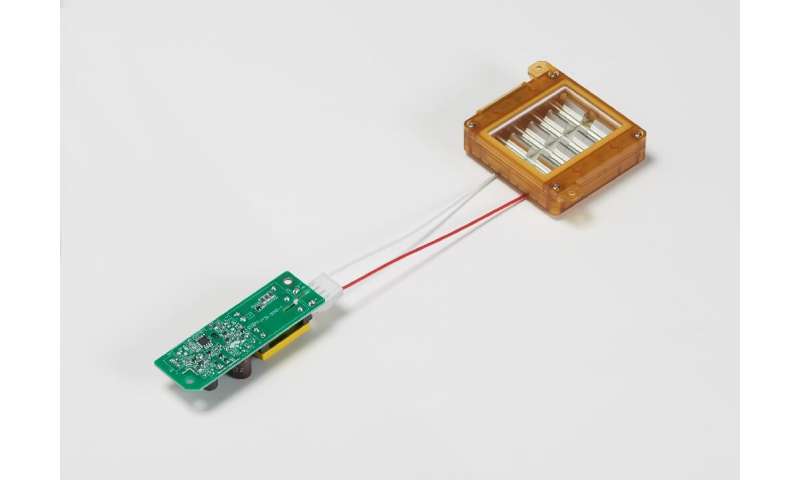
We used an excimer lamp based on a krypton-chlorine (Kr-Cl) gas mixture that emits principally at 222 nm. The lamp (High Current Electronics Institute, Tomsk, Russia) was air cooled with a 6,000-mm2 exit window (24). A custom bandpass filter (Omega Optical, Brattleboro, VT) was used to remove essentially all but the dominant 222-nm wavelength emission.
A UV spectrometer (Photon Control, BC, Canada) sensitive in the wavelength range from 200–360 nm was used to characterize the wavelength spectra emitted by the excimer lamp, and a deuterium lamp standard with a NIST-traceable spectral irradiance (Newport Corp, Stratford, CT) was used to calibrate the UV spectrometer.
Studies were also carried out with a conventional mercury germicidal lamp (Hygeaire, Atlantic Ultraviolet Corporation, Hauppauge, NY) with peak emission at 254 nm and used as positive control. An ILT1400 sensor equipped with a SEL220 detector (International Light Technologies, Peabody, MA) was used to measure the fluence rate from both lamps.
For acute exposures (MRSA and 3D tissues), the 222- and 254-nm lamps were positioned 9 and 99 cm, respectively, from the sample at which corresponded a power density of 0.036 mJ/cm2.
For chronic exposures of mice, delivery of 157 mJ/cm2 in a 7 h period was obtained by locating the 222- and the 254-nm lamps 41 and 205 cm from the samples, respectively; the corresponding power densities were 0.0062 mJ/cm2 for the 222-nm lamp and 0.0008 mJ/cm2 in the case of the conventional germicidal lamp.
Figure 1 shows the measured spectra emitted from the Kr-Cl excimer lamp that, together with the main excimer emission at 222 nm, also includes lower fluences of higher wavelength light (i.e. ~237 and ~258 nm).
To remove these more penetrating higher wavelengths and therefore potentially more harmful to human cells, we used a custom bandpass filter (Omega Optical, Brattleboro, VT). As shown in the inset in Fig. 1, the filtered lamp effectively emitted only the characteristic single wavelength of 222 nm.

References
1. Yin R, Dai T, Avci P, Jorge AE, de Melo WC, Vecchio D, et al. Light based anti-infectives: ultraviolet C irradiation, photodynamic therapy, blue light, and beyond. Curr Opin Pharmacol. 2013;13(5):731–62. [PMC free article] [PubMed] [Google Scholar]
2. McDevitt JJ, Rudnick SN, Radonovich LJ. Aerosol Susceptibility of Influenza Virus to UV-C Light. Appl Environ Microbiol. 2012;78(6):1666–9. [PMC free article] [PubMed] [Google Scholar]
3. Mitchell DL, Nairn RS. The (6-4) photoproduct and human skin cancer. Photo-dermatol. 1988;5(2):61–4. [PubMed] [Google Scholar]
4. Pfeifer GP, Besaratinia A. UV wavelength-dependent DNA damage and human non-melanoma and melanoma skin cancer. Photochem Photobiol Sci. 2012;11(1):90–7. [PMC free article] [PubMed] [Google Scholar]
5. Jose JG, Pitts DG. Wavelength dependency of cataracts in albino mice following chronic exposure. Exp Eye Res. 1985;41(4):545–63. [PubMed] [Google Scholar]
6. Soderberg PG. Acute cataract in the rat after exposure to radiation in the 300 nm wavelength region. A study of the macro-, micro- and ultrastructure. Acta Ophthalmol. 1988;66(2):141–52. [PubMed] [Google Scholar]
7. Buonanno M, Randers-Pehrson G, Bigelow AW, Trivedi S, Lowy FD, Spotnitz HM, et al. 207-nm UV light – A promising tool for safe low-cost reduction of surgical site infections. I: In vitro studies. PLoS ONE. doi:101371/journalpone0076968. eCollection 2013. [PMC free article] [PubMed] [Google Scholar]
8. Buonanno M, Stanislauskas M, Ponnaiya B, Bigelow AW, Randers-Pehrson G, Shuryak I, et al. 207-nm UV light – A promising tool for safe low-cost reduction of surgical site infections. II: In-vivo safety studies. PLoS One. 2016 Jun 8;11(6):e0138418. doi: 10.1371/journal.pone.0138418. [PMC free article] [PubMed] [CrossRef] [Google Scholar]
9. Sosnin EA, Oppenlander T, Tarasenko VF. Applications of capacitive and barrier discharge excilamps in photoscience. J Photochem Photobiol C: Photochem Rev. 2006;7:145–63. [Google Scholar]
10. Metzler DE, Metzler CM. Biochemistry: The Chemical Reactions of Living Cells. 2nd. San Diego: Academic Press; 2001. [Google Scholar]
11. Lorian V, Zak O, Suter J, Bruecher C. Staphylococci, in vitro and in vivo. Diagn Microbiol Infect Dis. 1985;3(5):433–44. [PubMed] [Google Scholar]
12. Goldfarb AR, Saidel LJ. Ultraviolet absorption spectra of proteins. Science. 1951;114(2954):156–7. [PubMed] [Google Scholar]
13. Setlow J. The molecular basis of biological effects of ultraviolet radiation and photoreactivation. In: Ebert M, Howard A, editors. Current topics in radiation research II. Amsterdam: North Holland Publishing Company; 1966. pp. 195–248. [Google Scholar]
14. Doughty MJ, Zaman ML. Human corneal thickness and its impact on intraocular pressure measures: a review and meta-analysis approach. Surv Ophthalmol. 2000;44(5):367–408. [PubMed] [Google Scholar]
15. Kolozsvari L, Nogradi A, Hopp B, Bor Z. UV absorbance of the human cornea in the 240- to 400-nm range. Invest Ophthalmol Vis Sci. 2002;43(7):2165–8. [PubMed] [Google Scholar]
16. Anderson DJ. Surgical site infections. Infect Dis Clin North Am. 2011;25(1):135–53. [PubMed] [Google Scholar]
17. Allegranzi B, Bischoff P, de Jonge S, Kubilay NZ, Zayed B, Gomes SM, et al. New WHO recommendations on preoperative measures for surgical site infection prevention: an evidence-based global perspective. Lancet Infect Dis. 2016 [PubMed] [Google Scholar]
18. Fry DE, Barie PS. The changing face of Staphylococcus aureus: a continuing surgical challenge. Surg Infect. 2011;12(3):191–203. [PubMed] [Google Scholar]
19. Anderson DJ, Kaye KS. Staphylococcal surgical site infections. Infect Dis Clin North Am. 2009;23(1):53–72. [PubMed] [Google Scholar]
20. Conner-Kerr TA, Sullivan PK, Gaillard J, Franklin ME, Jones RM. The effects of ultraviolet radiation on antibiotic-resistant bacteria in vitro. Ostomy Wound Manage. 1998;44(10):50–6. [PubMed] [Google Scholar]
21. Rao BK, Kumar P, Rao S, Gurung B. Bactericidal effect of ultraviolet C (UVC), direct and filtered through transparent plastic, on gram-positive cocci: an in vitro study. Ostomy Wound Manage. 2011;57(7):46–52. [PubMed] [Google Scholar]
22. Ritter MA, Olberding EM, Malinzak RA. Ultraviolet lighting during orthopaedic surgery and the rate of infection. J Bone Joint Surg Am. 2007;89(9):1935–40. [PubMed] [Google Scholar]
23. Berg M, Bergman BR, Hoborn J. Ultraviolet radiation compared to an ultra-clean air enclosure. Comparison of air bacteria counts in operating rooms. J Bone Joint Surg Br. 1991;73(5):811–5. [PubMed] [Google Scholar]
24. Sosnin EA, Avdeev SM, Kuznetzova EA, Lavrent’eva LV. A bacterial barrier-discharge KrBr Excilamp. Instr Experiment Tech. 2005;48:663–6. [Google Scholar]
25. Anderson DJ, Sexton DJ, Kanafani ZA, Auten G, Kaye KS. Severe surgical site infection in community hospitals: epidemiology, key procedures, and the changing prevalence of methicillin-resistant Staphylococcus aureus. Infect Control Hosp Epidemiol. 2007;28(9):1047–53. [PubMed] [Google Scholar]
26. Hidron AI, Edwards JR, Patel J, Horan TC, Sievert DM, Pollock DA, et al. NHSN annual update: antimicrobial-resistant pathogens associated with healthcare-associated infections: annual summary of data reported to the National Healthcare Safety Network at the Centers for Disease Control and Prevention, 2006-2007. Infect Control Hosp Epidemiol. 2008;29(11):996–1011. [PubMed] [Google Scholar]
27. Lessa FC, Mu Y, Ray SM, Dumyati G, Bulens S, Gorwitz RJ, et al. Impact of USA300 methicillin-resistant Staphylococcus aureus on clinical outcomes of patients with pneumonia or central line-associated bloodstream infections. Clin Infect Dis. 2012;55(2):232–41. [PubMed] [Google Scholar]
28. Kubilus J, Hayden PJ, Ayehunie S, Lamore SD, Servattalab C, Bellavance KL, et al. Full thickness epiderm: a dermal-epidermal skin model to study epithelial-mesenchymal interactions. Altern Lab Anim. 2004;32(Suppl 1A):75–82. [PubMed] [Google Scholar]
29. Bissett DL, Hannon DP, Orr TV. Wavelength dependence of histological, physical, and visible changes in chronically UV-irradiated hairless mouse skin. Photochem Photobiol. 1989;50(6):763–9. [PubMed] [Google Scholar]
30. Bissett DL, Hannon DP, Orr TV. An animal model of solar-aged skin: histological, physical, and visible changes in UV-irradiated hairless mouse skin. Photochem Photobiol. 1987;46(3):367–78. [PubMed] [Google Scholar]
31. Sato K, Sugibayashi K, Morimoto Y. Species differences in percutaneous absorption of nicorandil. J Pharm Sci. 1991;80(2):104–7. [PubMed] [Google Scholar]
32. Bronaugh RL, Stewart RF, Congdon ER. Methods for in vitro percutaneous absorption studies. II. Animal models for human skin. Toxicol Appl Pharmacol. 1982;62(3):481–8. [PubMed] [Google Scholar]
33. Russell LM, Wiedersberg S, Delgado-Charro MB. The determination of stratum corneum thickness: an alternative approach. Eur J Pharm Biopharm. 2008;69(3):861–70. [PMC free article] [PubMed] [Google Scholar]
34. van Diest PJ, Brugal G, Baak JP. Proliferation markers in tumours: interpretation and clinical value. J Clin Pathol. 1998;51(10):716–24. [PMC free article] [PubMed] [Google Scholar]
35. Scholzen T, Gerdes J. The Ki-67 protein: from the known and the unknown. J Cell Physiol. 2000;182(3):311–22. [PubMed] [Google Scholar]
36. Terui T, Okuyama R, Tagami H. Molecular events occurring behind ultraviolet-induced skin inflammation. Curr Opin Allergy Clin Immunol. 2001;1(5):461–7. [PubMed] [Google Scholar]
37. Horio T, Miyauchi H, Sindhvananda J, Soh H, Kurokawa I, Asada Y. The effect of ultraviolet (UVB and PUVA) radiation on the expression of epidermal keratins. Br Dermatol. 1993;128(1):10–5. [PubMed] [Google Scholar]
38. Sano T, Kume T, Fujimura T, Kawada H, Higuchi K, Iwamura M, et al. Long-term alteration in the expression of keratins 6 and 16 in the epidermis of mice after chronic UVB exposure. Arch Dermatol Res. 2009;301(3):227–37. [PubMed] [Google Scholar]
39. Green H, Boll J, Parrish JA, Kochevar IE, Oseroff AR. Cytotoxicity and mutagenicity of low intensity, 248 and 193 nm excimer laser radiation in mammalian cells. Cancer Res. 1987;47(2):410–3. [PubMed] [Google Scholar]
40. Coohill TP. Virus-cell interactions as probes for vacuum-ultraviolet radiation damage and repair. Photochem Photobiol. 1986;44(3):359–63. [PubMed] [Google Scholar]
41. Crolla RM, van der Laan L, Veen EJ, Hendriks Y, van Schendel C, Kluytmans J. Reduction of surgical site infections after implementation of a bundle of care. PLoS One. 2012;7(9):e44599. [PMC free article] [PubMed] [Google Scholar]
42. Lidwell OM, Lowbury EJ, Whyte W, Blowers R, Stanley SJ, Lowe D. Airborne contamination of wounds in joint replacement operations: the relationship to sepsis rates. J Hosp Infect. 1983;4(2):111–31. [PubMed] [Google Scholar]
43. Gosden PE, MacGowan AP, Bannister GC. Importance of air quality and related factors in the prevention of infection in orthopaedic implant surgery. J Hosp Infect. 1998;39(3):173–80. [PubMed] [Google Scholar]
44. Stocks GW, O’Connor DP, Self SD, Marcek GA, Thompson BL. Directed air flow to reduce airborne particulate and bacterial contamination in the surgical field during total hip arthroplasty. J Arthroplasty. 2011;26(5):771–6. [PubMed] [Google Scholar]
45. de Carvalho CC. Biofilms: recent developments on an old battle. Recent Pat Biotechnol. 2007;1(1):49–57. [PubMed] [Google Scholar]
46. Rahim K, Saleha S, Zhu X, Huo L, Basit A, Franco OL. Bacterial contribution in chronicity of wounds. Microb Ecol. 2016 [PubMed] [Google Scholar]
47. Uplekar M, Weil D, Lonnroth K, Jaramillo E, Lienhardt C, Dias HM, et al. WHO’s new end TB strategy. Lancet. 2015;385(9979):1799–801. [PubMed] [Google Scholar]
48. Reed NG. The history of ultraviolet germicidal irradiation for air disinfection. Public Health Rep. 2010;125(1):15–27. [PMC free article] [PubMed] [Google Scholar]
49. Nardell EA, Bucher SJ, Brickner PW, Wang C, Vincent RL, Becan-McBride K, et al. Safety of upper-room ultraviolet germicidal air disinfection for room occupants: results from the Tuberculosis Ultraviolet Shelter Study. Public Health Rep. 2008;123(1):52–60. [PMC free article] [PubMed] [Google Scholar]
50. Escombe AR, Moore DA, Gilman RH, Navincopa M, Ticona E, Mitchell B, et al. Upper-room ultraviolet light and negative air ionization to prevent tuberculosis transmission. PLoS Med. 2009;6(3):e43. [PMC free article] [PubMed] [Google Scholar]
51. Linnes JC, Rudnick SN, Hunt GM, McDevitt JJ, Nardell EA. Eggcrate UV: a whole ceiling upper-room ultraviolet germicidal irradiation system for air disinfection in occupied rooms. Indoor Air. 2014;24(2):116–24. [PubMed] [Google Scholar]
52. Wengraitis S, Reed NG. Ultraviolet spectral reflectance of ceiling tiles, and implications for the safe use of upper-room ultraviolet germicidal irradiation. Photochem Photobiol. 2012;88(6):1480–8. [PubMed] [Google Scholar]
53. Sliney D. Balancing the risk of eye irritation from UV-C with infection from bioaerosols. Photochem Photobiol. 2013;89(4):770–6. [PubMed] [Google Scholar]


