


 Extract (6-MSITC) in Healthy Older Adults")
: An In-Depth Exploration into its Thermogenic Role and Social Significance")


Researchers at the University of Maryland School of Medicine (UMSOM) and School of Pharmacy (UMSOP) have discovered new drug compounds to potentially treat the novel coronavirus that causes COVID-19.
The compounds disrupt the functioning of a protein complex inside human cells that the researchers discovered is critical for the replication and survival of coronaviruses.
This finding could lead to the development of new broad-spectrum antiviral drugs that target viruses such as influenza, Ebola and coronaviruses, according to a new study published today in the Proceedings of the National Academy of Sciences (PNAS) journal.
The protein complex, called SKI complex, is a group of human proteins that regulates various aspects of the normal functioning of a cell.
In the new study, the researchers discovered that this complex also plays a crucial role in helping a virus replicate its genetic material, called RNA, within the cells it infects.
“We determined that disrupting the SKI complex keeps the virus from copying itself, which essentially destroys it,” said study corresponding author Matthew Frieman, Ph.D., Associate Professor of Microbiology and Immunology at the UMSOM.
“We also identified compounds that targeted the SKI complex, not only inhibiting coronaviruses but also influenza viruses and filoviruses, such as the one that causes Ebola.”
He and his colleagues from the School of Pharmacy’s Computer-Aided Drug Design Center and the Center for Biomolecular Therapeutics at the UMSOM used computer modeling to identify a binding site on the SKI complex and identified chemical compounds that could bind to this site.
Subsequent experimental analysis showed these compounds to have antiviral activity against coronaviruses, influenza viruses, and filoviruses (such as Ebola). Researchers from the National Institute of Allergy and Infectious Diseases also participated in this study.
The study was funded by Emergent BioSolutions, a biopharmaceutical company based in Gaithersburg, MD.
“These findings present an important first step in identifying potential new antivirals that could be used to treat a broad number of deadly infectious diseases,” said study lead author Stuart Weston, Ph.D., a research fellow at the UMSOM. Such drugs have the potential to treat infectious disease associated with future pandemics.
Next steps include conducting animal studies to learn more about the safety and efficacy of these experimental compounds, which are not approved by the Food and Drug Administration.
In other research efforts funded by the federal government, Dr. Frieman and his team are rapidly testing hundreds of drugs, approved and marketed for other conditions, to see whether any can be repurposed to prevent or treat COVID-19.
“As we face a potentially long, hard winter with COVID-19, our researchers continue their sustained efforts to advance innovations,” said E. Albert Reece, MD, Ph.D., MBA, Executive Vice President for Medical Affairs, UM Baltimore, and the John Z. and Akiko K. Bowers Distinguished Professor and Dean, University of Maryland School of Medicine.
“Basic research remains a vital part of this effort to leave us prepared for the next global pandemic.”
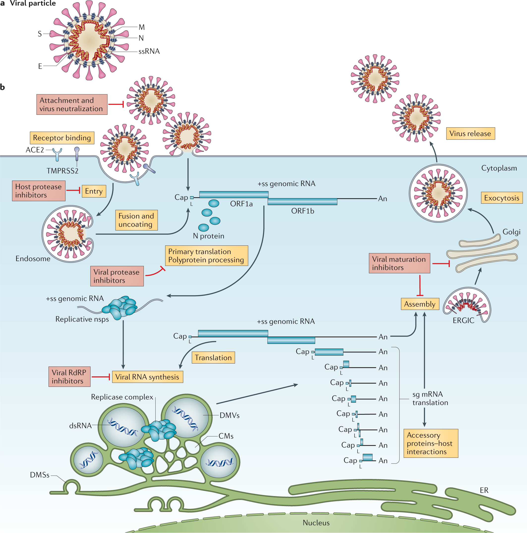
The initial steps of coronavirus infection involve the specific binding of the coronavirus spike (S) protein to the cellular entry receptors, which have been identified for several coronaviruses and include human aminopeptidase N (APN; HCoV-229E), angiotensin-converting enzyme 2 (ACE2; HCoV-NL63, SARS-CoV and SARS-CoV-2) and dipeptidyl peptidase 4 (DPP4; MERS-CoV).
The expression and tissue distribution of entry receptors consequently influence viral tropism and pathogenicity. During the intracellular life cycle (Fig. 1), coronaviruses express and replicate their genomic RNA to produce full-length copies that are incorporated into newly produced viral particles. Coronaviruses possess remarkably large RNA genomes flanked by 5′ and 3′ untranslated regions that contain cis-acting secondary RNA structures essential for RNA synthesis.
At the 5′ end, the genomic RNA features two large open reading frames (ORFs; ORF1a and ORF1b) that occupy two-thirds of the capped and polyadenylated genome. ORF1a and ORF1b encode 15–16 non-structural proteins (nsp), of which 15 compose the viral replication and transcription complex (RTC) that includes, amongst others, RNA-processing and RNA-modifying enzymes and an RNA proofreading function necessary for maintaining the integrity of the >30 kb coronavirus genome3. ORFs that encode structural proteins and interspersed ORFs that encode accessory proteins are transcribed from the 3′ one-third of the genome to form a nested set of subgenomic mRNAs (sg mRNAs).
Coronavirus accessory proteins are highly variable sets of virus-specific proteins that display limited conservation even within individual species but they are principally thought to contribute to modulating host responses to infection and are determinants of viral pathogenicity4,5.
Nevertheless, the molecular functions of many accessory proteins remain largely unknown owing to the lack of homologies to accessory proteins of other coronaviruses or to other known proteins6.
Despite the previous public health emergencies caused by the SARS-CoV and MERS-CoV outbreaks and the impact of the ongoing SARS-CoV-2 pandemic on society and human health, intervention strategies to combat coronavirus infections are only in their early stages and await proof of clinical efficacy.
Their development intimately relies on the deepened understanding of basic mechanisms of coronavirus gene functions as well as of the molecular interactions with host factors. Since the discovery of the first coronavirus (avian infectious bronchitis virus) in the 1930s7 and the discovery of the first human coronaviruses (HCoV-229E and HCoV-OC43) in the 1960s8,9, the coronavirus research field has made substantial progress in understanding the basic principles of coronavirus replication and pathogenesis (Box 1).
This advancement was accelerated after the emergence of SARS-CoV in 2002 and MERS-CoV in 2012 and has broadened our view on coronaviruses as zoonotic pathogens that can severely affect human health. Moreover, the unprecedented speed and technical progress of coronavirus research that has become evident in a few months after the appearance of SARS-CoV-2 at the end of 2019 has led to a rapidly growing understanding of this newly emerging pathogen and of its associated disease, COVID-19.
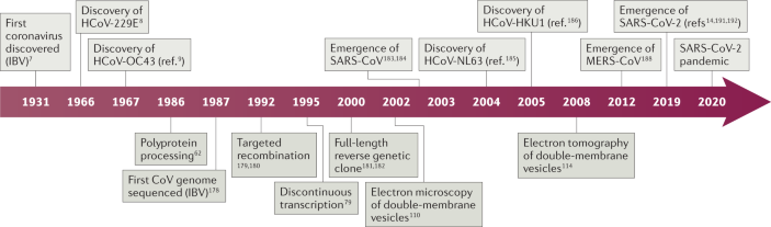
Box 1 Milestones in coronavirus discovery and research
Coronaviruses (CoVs) are a large family of viruses long known to infect a wide variety of mammalian and avian species, including livestock and companion animals. In 1931, the avian infectious bronchitis virus (IBV) was the first coronavirus to be discovered7. Later, in 1966 and 1967, the first human coronaviruses, HCoV-229E and HCoV-OC43, were discovered8,9.
The following period was essential in the discovery of research milestones that majorly contributed to coronavirus knowledge: polyprotein processing (1986)62, first full-length coronavirus genome sequence (1987)178, first recombinant coronaviruses engineered by targeted recombination (1992)179,180, discontinuous transcription (1995)78, full-length reverse genetic clones (2000, 2001)181,182 and electron microscopy of double-membrane vesicles (2002)110.
The zoonotic emergence of severe acute respiratory syndrome coronavirus (SARS-CoV) and the subsequent SARS epidemic in 2002–2003 caused 8,000 documented SARS cases, 10% of which had lethal consequences183,184. As human-to-human transmission mainly occurred after the onset of symptoms, drastic public health measures, including travel restrictions and isolation of infected patients, succeeded in containing the international spread to limited foyers of infections.
The SARS epidemic was followed by an increased amount of virus screening and sequencing, which led to the identification of HCoV-NL63 and HCoV-HKU1 (refs185,186). In contrast to SARS-CoV, HCoV-229E, HCoV-OC43, HCoV-NL63 and HCoV-HKU1 circulate annually and usually cause only mild upper respiratory tract symptoms in immunocompetent individuals185,186,187.
In 2008, SARS-CoV-induced double-membrane vesicles were first shown using electron tomography114. The emergence of a second highly pathogenic coronavirus of zoonotic origin, MERS-CoV, resulted in more than 2,500 human MERS cases since 2012, associated with virus-induced lung injuries and severe clinical manifestations (36% case fatality rate)188.
MERS-CoV also originated from bats and established an animal reservoir in dromedary camels189,190. Despite sporadic zoonotic transmissions to humans upon prolonged contact and the limited human-to-human transmission, MERS-CoV infections are still detected190.
Recently, the pathogenic SARS-CoV-2 rapidly spread in the human population after a likely spillover from bats or from a yet unidentified intermediate host14,191,192.
As of October 2020, more than 40 million COVID-19 cases have been declared in over 200 countries, causing more than 1 million deaths (COVID-19 Dashboard).
SARS-CoV-2 targets both upper and lower respiratory tract tissues and efficient human-to-human transmission occurs even before the onset of symptoms36,193. Clinical manifestations range from asymptomatic or mild infections to acute lung inflammation and pneumonia, mostly in the elderly and patients with comorbidities36,194,195.
reference link: https://www.nature.com/articles/s41579-020-00468-6
The SKI complex, a protein complex involved in various RNA metabolism pathways, has been identified as a potential target for broad-spectrum antiviral drugs. Already, chemicals have been identified that interact with the SKI complex. Encouragingly, these chemicals have been shown to prevent the replication of coronaviruses, influenza viruses, and filoviruses in mammalian cell culture.
The SKI complex was originally identified in yeast, but it is also found in mammals. Both these SKI contexts figured in a recent search for broad-spectrum, host-directed, antiviral targets. The search, conducted by scientists at the University of Maryland School of Medicine (UMSOM), began with yeast suppressor screening, the intent of which was to find a functional genetic interaction between viral proteins and eukaryotic proteins.
Screens were conducted with influenza A virus (IAV) and Middle East respiratory syndrome coronavirus (MERS-CoV). The eukaryotic proteins of interest were those potentially involved in supporting viral replication and survival.
The screens suggested that the SKI was needed by both viruses.
Next, conducting experiments in mammalian systems, the UMSOM scientists determined that siRNA-mediated knockdown of SKI genes inhibited replication of IAV and MERS-CoV. This finding prompted the UMSOM scientists to consider how the SKI complex could be targeted by potential antiviral agents.
Details of this work appeared November 12 in the Proceedings of the National Academy of Sciences (PNAS), in an article entitled, “The SKI complex is a broad-spectrum, host-directed antiviral drug target for coronaviruses, influenza, and filoviruses.”
“In silico modeling and database screening identified a binding pocket on the SKI complex and compounds predicted to bind,” the article’s authors wrote. “Experimental assays of those compounds identified three chemical structures that were antiviral against IAV and MERS-CoV along with the filoviruses Ebola and Marburg and two further coronaviruses, SARS-CoV and SARS-CoV-2.”
The chemicals’ antiviral activity, the authors suggested, occurs through inhibition of viral RNA production.
“We determined that disrupting the SKI complex keeps the virus from copying itself, which essentially destroys it,” said Matthew Frieman, PhD, the study’s corresponding author and associate professor of microbiology and immunology at the UMSOM. “We also identified compounds that targeted the SKI complex, not only inhibiting coronaviruses but also influenza viruses and filoviruses, such as the one that causes Ebola.”
He and his colleagues from the School of Pharmacy’s Computer-Aided Drug Design Center and the Center for Biomolecular Therapeutics at the UMSOM used computer modeling to identify a binding site on the SKI complex and identified chemical compounds that could bind to this site.
Subsequent experimental analysis showed these compounds to have antiviral activity against coronaviruses, influenza viruses, and filoviruses (such as Ebola). Researchers from the National Institute of Allergy and Infectious Diseases also participated in this study.
The study was funded by Emergent BioSolutions, a biopharmaceutical company based in Gaithersburg, MD.
“These findings present an important first step in identifying potential new antivirals that could be used to treat a broad number of deadly infectious diseases,” said Stuart Weston, PhD, the study’s lead author and a research fellow at the UMSOM. Such drugs have the potential to treat infectious disease associated with future pandemics.
Next steps include conducting animal studies to learn more about the safety and efficacy of these experimental compounds, which are not approved by the U.S. Food and Drug Administration.
“At this stage, our compounds that display broad antiviral activity are only modeled to interreact with the SKI complex,” the article’s authors noted. “We find that loss of the SKI complex through siRNA targeting removes the antiviral activity of [one of our compounds], suggesting a requirement for the complex to produce the observed antiviral activity.
Future work is aimed at further analyzing whether the compounds bind directly to the SKI complex using structural biology approaches and investigating whether other roles of the SKI complex are impacted.”
| NCBI Gene | 6497 |
|---|---|
| Symbol | SKI |
| Taxonomy | Homo sapiens (human) |
| Dates | Modify2020-11-09Create2016-09-14 |
| The SKI gene provides instructions for making a protein involved in a signaling pathway that transmits chemical signals from the cell surface to the nucleus. This pathway, called the transforming growth factor beta (TGF-β) pathway, allows the environment outside the cell to affect how the cell produces other proteins. It helps regulate cell growth and division (proliferation), the process by which cells mature to carry out special functions (differentiation), cell movement (motility), and the self-destruction of cells (apoptosis). Through this pathway, a group of proteins called the SMAD complex is turned on (activated). The activated SMAD protein complex moves to the cell nucleus and attaches (binds) to specific areas of DNA to control the activity of particular genes, which help regulate various cellular processes. The SKI protein controls the activity of the TGF-β pathway by binding to certain SMAD proteins, which interrupts signaling through the pathway. SKI protein binding within the cell can keep the SMAD protein complex from entering the nucleus, so it is unable to activate genes. Binding of the SKI protein can also occur in the nucleus. Although the SMAD complex binds to DNA, the SKI protein attracts other proteins (corepressors) that block its ability to turn genes on.The SKI protein is found in many cell types throughout the body and appears to play a role in the development of many tissues, including the skull, other bones, skin, and brain. |
This gene encodes the nuclear protooncogene protein homolog of avian sarcoma viral (v-ski) oncogene. It functions as a repressor of TGF-beta signaling, and may play a role in neural tube development and muscle differentiation. [provided by RefSeq, Oct 2009]
The v-Ski avian retroviral oncogene is postulated to act as a transcription factor. Since protein-protein interactions have been shown to play an important role in the transcription process, we attempted to identify Ski protein partners with the yeast two-hybrid system.
Using v-Ski sequence as bait, the human gene skip (Ski Interacting Protein) was identified as encoding a protein which interacts with both the cellular and viral forms of Ski in the two-hybrid system.
Skip is highly homologous to the Drosophila melanogaster protein Bx42 which is found associated with chromatin in transcriptionally active puffs of salivary glands. The Ski-Skip interaction is potentially important in Ski’s transforming activity since Skip was demonstrated to interact with a highly conserved region of Ski required for transforming activity. Like Ski, Skip is expressed in multiple tissue types and is localized to the cell nucleus.
More information: Stuart Weston et al, The SKI complex is a broad-spectrum, host-directed antiviral drug target for coronaviruses, influenza, and filoviruses, Proceedings of the National Academy of Sciences (2020). DOI: 10.1073/pnas.2012939117


[…] COVID-19: disrupting the SKI protein complex keeps the virus from copying… […]
[…] COVID-19: disrupting the SKI protein complex keeps the virus from copying… […]