


 Extract (6-MSITC) in Healthy Older Adults")
: An In-Depth Exploration into its Thermogenic Role and Social Significance")


Stimulating humans’ sense of smell to prevent conditions such as Alzheimer’s Disease is the focus of international research led by the University of Otago.
The olfactory system, or sense of smell, is known to be dysfunctional in the early stages of Alzheimer’s and Parkinson’s disease. It is also shown that proper olfactory function can play a key role in regaining consciousness after brain injuries.
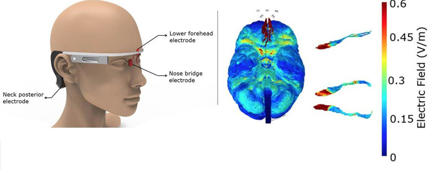
The Otago research centers around a wearable concept prototype, similar to Google-glasses, which produces small electronic pulses on the skin to stimulate the olfactory nervous system.
Areas of the brain prone to Alzheimer’s, Parkinson’s, and coma, can be jump-started to reduce or reverse the onset of those serious conditions.
Lead author, Associate Professor Yusuf Ozgur Cakmak from Otago’s Department of Anatomy, says promising early results pave the way for developing the world’s first non-invasive, wearable electrical stimulation system to target the olfactory regions.
“Olfactory nerves have terminals deep in the brain regions which influence memory and navigation. We’re hopeful this method will help stimulate these networks to alleviate symptoms or suppress the progression of Alzheimer’s disease to Dementia. It also has potential to help coma recovery and Parkinson’s disease,” Associate Professor Cakmak says.
Modulation of the olfactory regions has been attempted successfully with electrical stimulation previously, either directly (intraoperatively through the nasal bones) or indirectly through the vagus nerve. This research sought to develop a means of delivering electrical stimulation to the olfactory region in a non-invasive fashion and in a way that is simpler, easier, and less cumbersome.
“Applying this treatment via a headset on a hair-free zone that can be worn in daily routine instead of more invasive treatments makes this method unique ” Associate Professor Cakmak adds.
The multiple electrode configurations have been tested with the aid of electrical field modelling that is validated with direct human brain recordings during brain surgery.
Otago’s research team is collaborating with New York based company Soterix Medical, a world leader in non-invasive neuromodulation and brain monitoring technology. The international team will be testing their wearable stimulator in a clinical trial in 2020.
The study, “Optimized Electrode Placements for Non-Invasive Electrical Stimulation of the Olfactory Bulb and Olfactory Mucosa,” has been published in the specialist journal Frontiers in Neuroscience.
One in 10 people age 65 and older (10%) has Alzheimer’s dementia and 13.8 million people in the US age 65 and older are projected to have Alzheimer’s dementia by 2050, according to the Alzheimer’s Association.
Olfaction is the chemical sensation of gaseous odorants colloquially referred to as the ability to smell. The olfactory nerve (cranial nerve one) in coordination with other neuroanatomical structures in the nasal passages, neurotransmitters, and the cerebral cortex is responsible for carrying out this intricate chemosensory process.
In humans, olfaction closely couples to other complex functions such as gustation (taste) and involuntary memory formation.[1][2] From an evolutionary standpoint, an intact sense of smell is critical for evaluating the safety of ingestible substances, assessing impending danger, and recognizing social relationships.
The ability to perceive and detect orders tends to decline with normal aging.[3] In a clinical setting, changes in olfaction may represent the initial presentation of the underlying pathology and warrant a thorough medical evaluation.
Issues of Concern
Anosmia refers to the complete inability to detect odorants. The underlying causes of anosmia are highly variable and will be discussed in further detail. According to the National Institute on Deafness and Other Communication Disorders, approximately 1.4% of the population experiences dysfunction of the olfactory system along with some degree of concomitant loss of smell.[4]
Cellular
The olfactory system is at the roof of the nasal cavity at the cribriform plate – a perforated portion of the ethmoid bone separating the frontal lobe of the cerebrum from the nasal cavity. Odorant molecules within the nasal passages first encounter receptors on the primary cilia of olfactory sensory neurons.
Each neuron expresses a single type of protein receptor on these dendritic extensions. However, individual odorants can bind to many different receptor proteins. The dendritic ends of these first-order neurons are within a thin layer of mucus with adjacent supporting epithelium. Bowman glands secrete serous fluid rich in glycoprotein, which warms, moistens, and traps air, helping dissolve gaseous odorant particles.
The axonal components of individual olfactory sensory neurons then combine to form neurovascular bundles that project through the cribriform plate. These collective bundles of axons form the olfactory nerves. Axonal projections of olfactory nerves synapse with the dendrites of mitral and tufted cells in spherical structures known as glomeruli. Glomeruli are found on the surface of the olfactory bulb and are critical structures for transducing olfaction. Each glomerulus receives converging axons from olfactory neurons that express the same specific protein receptors.
Humans are estimated to have 1100 to 1200 glomeruli within each olfactory bulb.[5] Second-order mitral cells then project via olfactory tracts to specific areas within the brain that process olfactory information, including the piriform cortex, olfactory tubercle, amygdala, and entorhinal cortex.[6][7][8]
Development
Evidence of olfaction has been demonstrated in fetuses as early as 30 weeks’ gestation characterized by discriminative responses to odorous molecules in amniotic fluid.[9] Also, it is at this gestational age that olfactory bulbs are visible on magnetic resonance imaging (MRI). Olfactory bulbs continue undergoing neurodevelopmental and maturational changes until approximately two years of age.[10]
Kallmann syndrome is a congenital disorder characterized by hypogonadotropic hypogonadism along with anosmia due to bilateral agenesis or hypoplasia of the olfactory bulbs.[11] Acquired abnormalities of olfaction secondary to perinatal toxic insult are also well documented. A study of pregnant mice drinking 10% ethanol demonstrated hypoplastic olfactory bulbs and impaired odor discrimination in the offspring persisting into adulthood.[12]
Organ Systems Involved
The nose contains the olfactory organs at its superior pole while simultaneously serving its function in the respiratory system. Inside the anterior portion of each nostril is the nasal vestibule, a cartilaginous structure lined with squamous epithelium and small hairs called vibrissae. These hairs filter dust and other particles from inspired air.[13] Within each nasal cavity are three thin, shelf-shaped spongy bones originating from the lateral walls referred to as nasal concha (superior, middle, and inferior), also called turbinates. These increase the surface area of the nasal cavities and are highly vascularized to help condition air by rapid warming and humidification. The superior concha contains the olfactory organs.[14]
The peripheral components of the olfactory system, such as the olfactory sensory neurons, nerves, bulbs, and tracts, are organized structures within the central nervous system. Finally, the central processing of chemosensory input within various brain structures remains a highly complex process under current scientific investigation.[15]
Function
The olfactory system serves multiple functions in humans. Through direct connection with the limbic system and cerebral cortex, smells intertwine with experiencing emotions and memories. Olfaction also serves a role in shaping behaviors and communication between animals.[16] Odorants within the environment provide fundamental information for survival. Several species rely on olfaction to identify nutritional resources, mates, toxins, predators, and impending danger.[17]
Mechanism
The process of olfaction involves the conversion of a chemical stimulus, an odorant, into an electrical signal sent to the brain for interpretation. This mechanism begins after olfactory sensory neurons depolarize in response to the binding of an odorant molecule to G-protein coupled receptors (GPCR). The dissociated G protein activates an intracellular cascade via adenylyl cyclase producing a molecule of cyclic adenosine monophosphate (cAMP) that binds and opens ion channels within the neuron’s plasma membrane. Subsequently, an influx of positive sodium and calcium ions and an efflux of negative chloride ions occurs. Neuronal depolarization continues until the threshold potential occurs, firing a resulting action potential. The action potential travels down the olfactory nerves through the cribriform plate towards glomeruli in the olfactory bulb. The glomeruli then project to specific areas within the brain where higher-level processing, modulation, and interpretation occur.[18]
Related Testing
Testing the olfactory nerve is an essential component when conducting a comprehensive cranial nerve examination. The examiner can instruct the patient to occlude one nare while holding a discretely scented object (coffee grounds, orange peels, or tobacco are common choices) and asking the patient to identify the smell. Note, using an alcohol swab should be avoided because the nociceptive intranasal odorants may also stimulate the chemoreceptors of the trigeminal nerve (cranial nerve five) and bypass or interfere with the olfactory system.[19]
Pathophysiology
The pathophysiology of olfactory disorders can be better understood by first identifying the type of dysfunction present. Anosmia exists on a spectrum ranging from a complete lack of smell to a diminished ability or hyposmia (partial anosmia). Furthermore, a disturbance in olfaction may present as the inability to distinguish odorants apart from one another, known as olfactory agnosia. Distortions or pathologic alterations in perception can occur, collectively referred to as dysosmias.
Interestingly, spontaneous experience of smell in the absence of an odorant stimulus can occur. Such chemosensory hallucinations are designated phantosmias. Reports exist of the presence of phantosmias as high as 55% in transient epileptic amnesia.[20] Clinically, it is essential to distinguish the laterality (unilateral or bilateral) anytime an abnormality in olfaction is encountered to investigate the underlying cause further.[21]
Alterations in the ability to smell can be the result of disease processes, environmental exposure, or simply a product of normal aging. In adults under 65 years of age, the estimated prevalence of olfactory dysfunction is approximately 2%. However, this number increases drastically to 75% in populations over 80 years old.[22] These changes are likely multifactorial, caused in part by the ossification of the cribriform plate and a reduction in the size of its foramina.[23] Additionally, the cumulative damage to olfactory receptors encountered throughout one’s lifetime appears to play a role in the age-related olfactory decline.[3]
Odor Fear Conditioning in Humans
During the past ten years, several studies using the functional magnetic resonance imaging (fMRI) technique, have investigated the networks involved in fear conditioning in humans (Cheng et al. 2003; Buchel and Dolan 2000; LaBar et al. 1998). Most of these studies suggest that the amygdala is part of the circuit, thus corroborating the data from the animal literature. Interestingly, in a recent work, Li et al. (2008) used an odor fear conditioning paradigm in humans in order to investigate how aversive learning enhances perceptual acuity of sensory signal. During conditioning, the CS odor presentation coterminated with electric shock (US), whereas presentation of its chiral counterpart (enantiomer) was not associated with the US.
The authors combined multivariate fMRI with olfactory psychophysics, and hereby showed that initially indistinguishable odor enantiomers become discriminable after aversive conditioning. In parallel, fMRI data demonstrated progressive decreases in amygdala activity evoked by the learned aversive odor as learning proceeded, together with increases in the orbitofrontal cortex. Interestingly, the authors also measured changes in activity in the piriform cortex.
They reported that spatial patterns of fMRI activity in the posterior piriform cortex between the two enantiomers were highly correlated before conditioning, but became more distinct after conditioning. This effect was specific to the posterior piriform cortex, as it was not observed in the anterior piriform cortex.
These findings confirm and extend previous data reported by the same group, showing a double dissociation in the piriform cortex, whereby posterior regions encode quality, and anterior regions encode structure (Gottfried et al. 2006; Li et al. 2006). Taken together, these data indicate that aversive learning induces plasticity in the posterior piriform cortex that correlates with gains in odor enantiomer discrimination. This led the authors to propose that fear conditioning has the capacity to update perceptual representation of predictive cues, in addition to its well-recognized role in the acquisition of conditioned responses.
The data obtained in human odor fear conditioning are strikingly similar to those described in rats and suggest that whereas the amygdala plays a crucial role, a broad network of structures is involved in the learning, among which the piriform cortex seems to endorse a privileged status.
Clinical Significance
A benign decline in olfactory sensation is commonly encountered in aging populations and may appear transiently in the setting of intranasal inflammation or obstruction. However, when clinicians encounter abnormalities such as anosmia or hyposmia in a clinical setting, it is crucial to investigate the underlying etiology further. This process begins by taking a focused patient history and conducting a thorough neurologic examination, including evaluation of cranial nerve one.
A wide variety of diseases impact the olfactory function. Upper respiratory viral infections are the most common cause of both permanent anosmia and hyposmia.[24] Neurotropic viruses can cause permanent damage to the neural tissue of the olfactory system. In the setting of sinusitis, inflamed nasal mucosa and increased mucus production may result in nasal obstruction and commonly causes transient and incomplete anosmia.[25] This condition is one of the common causes of transient anosmia, frequently seen in all age groups.
Recently, studies have identified anosmia as a vital early sign of neurodegenerative disorders such as Parkinson and Alzheimer disease.[26][27] So it is crucial to check the olfactory function in the evaluation of the neurodegenerative diseases. Facial trauma, especially fractures involving injury to the cribriform plate, can also precipitate a loss of smell.[28] During the facial trauma, there are sometimes a fracture of the cribriform plate and the release of the CSF fluid. So, we have to do thorough work up in these cases to avoid potential infection to the brain and meninges.
Similarly, meningiomas of the olfactory groove and other intracranial masses can be secondary causes of olfaction loss.[29] Scientific evidence also supports a strong association between olfactory dysfunction and schizophrenia.[30] Current investigations of olfactory stimuli inciting migraine headaches and hyperemesis gravidarum in pregnancy suggest a common mechanism involving allelic variations in dopaminergic receptors.[31] Anecdotally, women have reported hyperosmia during pregnancy, but the scientific understanding of this phenomenon remains limited.[32]
Roughly 5 to 7% of patients with head injuries also reported a decrease in the olfactory sense. Some patients with diabetes also have dysfunction of the olfactory nerve. Olfactory hallucinations also occur in hippocampal lesions or psychosis. Patients describe olfactory hallucinations as unpleasant and strange odors.
References
- 1.Bruch RC, Kalinoski DL, Kare MR. Biochemistry of vertebrate olfaction and taste. Annu Rev Nutr. 1988;8:21-42. [PubMed]
- 2.Aso Y, Rubin GM. Dopaminergic neurons write and update memories with cell-type-specific rules. Elife. 2016 Jul 21;5 [PMC free article] [PubMed]
- 3.Attems J, Walker L, Jellinger KA. Olfaction and Aging: A Mini-Review. Gerontology. 2015;61(6):485-90. [PubMed]
- 4.Scangas GA, Bleier BS. Anosmia: Differential diagnosis, evaluation, and management. Am J Rhinol Allergy. 2017 Jan 01;31(1):3-7. [PubMed]
- 5.Pinching AJ, Powell TP. The neuropil of the glomeruli of the olfactory bulb. J Cell Sci. 1971 Sep;9(2):347-77. [PubMed]
- 6.Persaud KC. Engineering Aspects of Olfaction. In: Persaud KC, Marco S, Gutiérrez-Gálvez A, editors. Neuromorphic Olfaction. CRC Press/Taylor & Francis; Boca Raton (FL): 2013. [PubMed]
- 7.Hatt H. Molecular and cellular basis of human olfaction. Chem Biodivers. 2004 Dec;1(12):1857-69. [PubMed]
- 8.Benignus VA, Prah JD. Olfaction: anatomy, physiology and behavior. Environ Health Perspect. 1982 Apr;44:15-21. [PMC free article] [PubMed]
- 9.Sarnat HB, Flores-Sarnat L, Wei XC. Olfactory Development, Part 1: Function, From Fetal Perception to Adult Wine-Tasting. J Child Neurol. 2017 May;32(6):566-578. [PubMed]
- 10.Schneider JF, Floemer F. Maturation of the olfactory bulbs: MR imaging findings. AJNR Am J Neuroradiol. 2009 Jun;30(6):1149-52. [PMC free article] [PubMed]
- 11.Shetty S, Kapoor N, John RA, Paul TV. Olfactory Agenesis in Kallmann Syndrome (KS). J Clin Diagn Res. 2015 Apr;9(4):OJ01. [PMC free article] [PubMed]
- 12.Akers KG, Kushner SA, Leslie AT, Clarke L, van der Kooy D, Lerch JP, Frankland PW. Fetal alcohol exposure leads to abnormal olfactory bulb development and impaired odor discrimination in adult mice. Mol Brain. 2011 Jul 07;4:29. [PMC free article] [PubMed]
- 13.Stoddard DG, Pallanch JF, Hamilton GS. The effect of vibrissae on subjective and objective measures of nasal obstruction. Am J Rhinol Allergy. 2015 Sep-Oct;29(5):373-7. [PubMed]
- 14.Freeman SC, Karp DA, Kahwaji CI. StatPearls [Internet]. StatPearls Publishing; Treasure Island (FL): Jul 10, 2020. Physiology, Nasal. [PubMed]
- 15.Benjaminsson S, Herman P, Lansner A. Performance of a Computational Model of the Mammalian Olfactory System. In: Persaud KC, Marco S, Gutiérrez-Gálvez A, editors. Neuromorphic Olfaction. CRC Press/Taylor & Francis; Boca Raton (FL): 2013. [PubMed]
- 16.Zarzo M. The sense of smell: molecular basis of odorant recognition. Biol Rev Camb Philos Soc. 2007 Aug;82(3):455-79. [PubMed]
- 17.Hoover KC. Smell with inspiration: the evolutionary significance of olfaction. Am J Phys Anthropol. 2010;143 Suppl 51:63-74. [PubMed]
- 18.Pinto JM. Olfaction. Proc Am Thorac Soc. 2011 Mar;8(1):46-52. [PMC free article] [PubMed]
- 19.Brand G. Olfactory/trigeminal interactions in nasal chemoreception. Neurosci Biobehav Rev. 2006;30(7):908-17. [PubMed]
- 20.Savage SA, Butler CR, Milton F, Han Y, Zeman AZ. On the nose: Olfactory disturbances in patients with transient epileptic amnesia. Epilepsy Behav. 2017 Jan;66:113-119. [PMC free article] [PubMed]
- 21.Doty RL. The olfactory system and its disorders. Semin Neurol. 2009 Feb;29(1):74-81. [PubMed]
- 22.Doty RL, Shaman P, Dann M. Development of the University of Pennsylvania Smell Identification Test: a standardized microencapsulated test of olfactory function. Physiol Behav. 1984 Mar;32(3):489-502. [PubMed]
- 23.Kalmey JK, Thewissen JG, Dluzen DE. Age-related size reduction of foramina in the cribriform plate. Anat Rec. 1998 Jul;251(3):326-9. [PubMed]
- 24.Betchen SA, Doty RL. Bilateral detection thresholds in dextrals and sinistrals reflect the more sensitive side of the nose, which is not lateralized. Chem Senses. 1998 Aug;23(4):453-7. [PubMed]
- 25.Raviv JR, Kern RC. Chronic sinusitis and olfactory dysfunction. Otolaryngol Clin North Am. 2004 Dec;37(6):1143-57, v-vi. [PubMed]
- 26.Hüttenbrink KB, Hummel T, Berg D, Gasser T, Hähner A. Olfactory dysfunction: common in later life and early warning of neurodegenerative disease. Dtsch Arztebl Int. 2013 Jan;110(1-2):1-7, e1. [PMC free article] [PubMed]
- 27.Hawkes C. Olfaction in neurodegenerative disorder. Mov Disord. 2003 Apr;18(4):364-72. [PubMed]
- 28.Reiter ER, DiNardo LJ, Costanzo RM. Effects of head injury on olfaction and taste. Otolaryngol Clin North Am. 2004 Dec;37(6):1167-84. [PubMed]
- 29.Jang WY, Jung S, Jung TY, Moon KS, Kim IY. Preservation of olfaction in surgery of olfactory groove meningiomas. Clin Neurol Neurosurg. 2013 Aug;115(8):1288-92. [PubMed]
- 30.Kiparizoska S, Ikuta T. Disrupted Olfactory Integration in Schizophrenia: Functional Connectivity Study. Int J Neuropsychopharmacol. 2017 Sep 01;20(9):740-746. [PMC free article] [PubMed]
- 31.Heinrichs L. Linking olfaction with nausea and vomiting of pregnancy, recurrent abortion, hyperemesis gravidarum, and migraine headache. Am J Obstet Gynecol. 2002 May;186(5 Suppl Understanding):S215-9. [PubMed]
- 32.Cameron EL. Pregnancy and olfaction: a review. Front Psychol. 2014;5:67. [PMC free article] [PubMed]
More information: Yusuf Ozgur Cakmak et al. Optimized Electrode Placements for Non-invasive Electrical Stimulation of the Olfactory Bulb and Olfactory Mucosa, Frontiers in Neuroscience (2020). DOI: 10.3389/fnins.2020.581503


[…] Alzheimer’s disease: the new wearable electrical stimulator can reduce or reverse… […]