


 Extract (6-MSITC) in Healthy Older Adults")
: An In-Depth Exploration into its Thermogenic Role and Social Significance")


Researchers at Umeå University in Sweden have for the first time at the atomic level succeeded in mapping what a virus looks like that causes diarrhea and annually kills about 50,000 children in the world.
The discovery may in the long run provide the opportunity for completely new types of treatments for other viral diseases such as COVID-19.
“The findings provide an increased understanding of how the virus gets through the stomach and intestinal system. Continued research can provide answers to whether this property can also be used to create vaccines that ride ‘free rides’ and thus be given in edible form instead of as syringes,” says Lars-Anders Carlson, researcher at Umeå University.
The virus that the researchers have studied is a so-called enteric adenovirus. It has recently been clarified that enteric adenoviruses are one of the most important factors behind diarrhea among infants, and they are estimated to kill more than 50,000 children under the age of five each year, mainly in developing countries.
Most adenoviruses are respiratory, that is, they cause respiratory disease, while the lesser-known enteric variants of adenovirus instead cause gastrointestinal disease.
The enteric adenoviruses therefore need to be equipped to pass through the acidic environment of the stomach without being broken down, so that they can then infect the intestines.
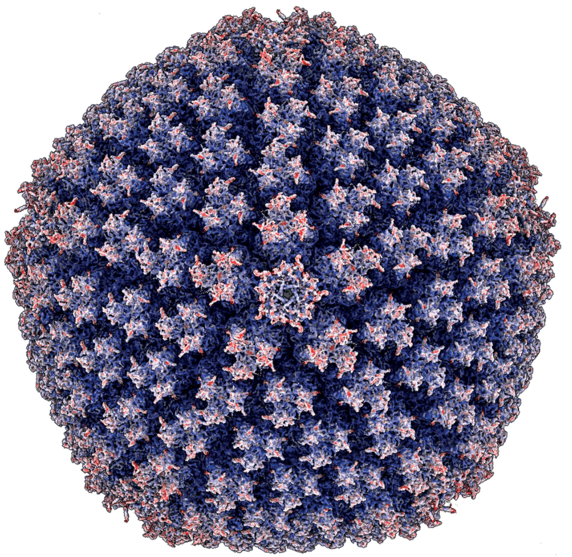
With the help of the advanced cryo-electron microscope available in Umeå, the researchers have now managed to take such detailed images of an enteric adenovirus that it has been possible to put a three-dimensional puzzle that shows what the virus looks like right down to the atomic level.
The virus is one of the most complex biological structures studied at this level. The shell that protects the virus’ genome when it is spread between humans consists of two thousand protein molecules with a total of six million atoms.
The researchers were able to see that the enteric adenovirus manages to keep its structure basically unchanged at the low pH value found in the stomach.
They could also see other differences compared to respiratory adenoviruses in how a particular protein is altered in the shell of the virus as well as new clues to how the virus packs its genome inside the shell. All in all, it provides an increased understanding of how the virus manages to move on to create disease and death.
“The hope is that you will be able to turn the ability that this unpleasant virus has to get to something that can instead be used as a tool to fight disease, perhaps even COVID-19. This is a step in the right direction, but it is still a long way off,” says Lars-Anders Carlson.
Several of the new vaccines being tested against COVID-19 are based on genetically modified adenovirus. Today, these adenovirus-based vaccines must be injected to work in the body.
If a vaccine could instead be based on enteric adenovirus, the vaccine might be given in edible form. This would, of course, facilitate large-scale vaccination.
The virus that the researchers have studied is called HAdV-F41. The study is published in the scientific journal Science Advances. It is a collaboration between Lars-Anders Carlson’s and Niklas Arnberg’s research groups at Umeå University.
Adenoviruses (AdVs) are common pathogens in human, causing diseases in airways, eyes, and intestine, but also in the liver, urinary tract, and/or adenoids1. To date, more than 100 human adenovirus (HAdV) types have been isolated, characterized, and classified into seven species (A-G)2.
The two sole members of HAdV species F, HAdV-F40 and F41, stand out amongst HAdVs due to their pronounced gastrointestinal tropism. These so-called enteric adenoviruses are a leading cause of diarrhoea and diarrhoea-associated mortality3 in young children, inferior only to Shigella and rotavirus4.
Diarrhoea is estimated to cause ∼530,000 deaths/year in children younger than five years world-wide5 and thus, there is a strong incentive to understand the structural and molecular basis of enteric HAdVs.
AdVs are double-stranded DNA viruses with an approximately ∼35 kbp genome sheltered in a large (∼950 Å in diameter), non-enveloped capsid with icosahedral symmetry6-11. At each of the twelve capsid vertices, penton base subunits organise as homopentamers12, anchoring the N-terminal tails of the protruding, trimeric fibres13,14 to the capsid.
Another so-called major capsid protein is the hexon protein15, present in 240 trimers per virion. Hexons are the main structural component of the virion facets and are organised to give the virion a pseudo T=25 symmetry (Fig. 1A). Hexon assemblies are stabilised by minor capsid protein IIIa (pIIIa), pVI and pVIII (located in the capsid interior) and by pIX (exposed on the capsid exterior)7,8,16.
To date, two human adenoviruses, HAdV-C59,10,17,18 and HAdV-D2619, but also individual capsid proteins or their subdomains12,15,20 of multiple adenovirus types, have been structurally determined at high resolution.
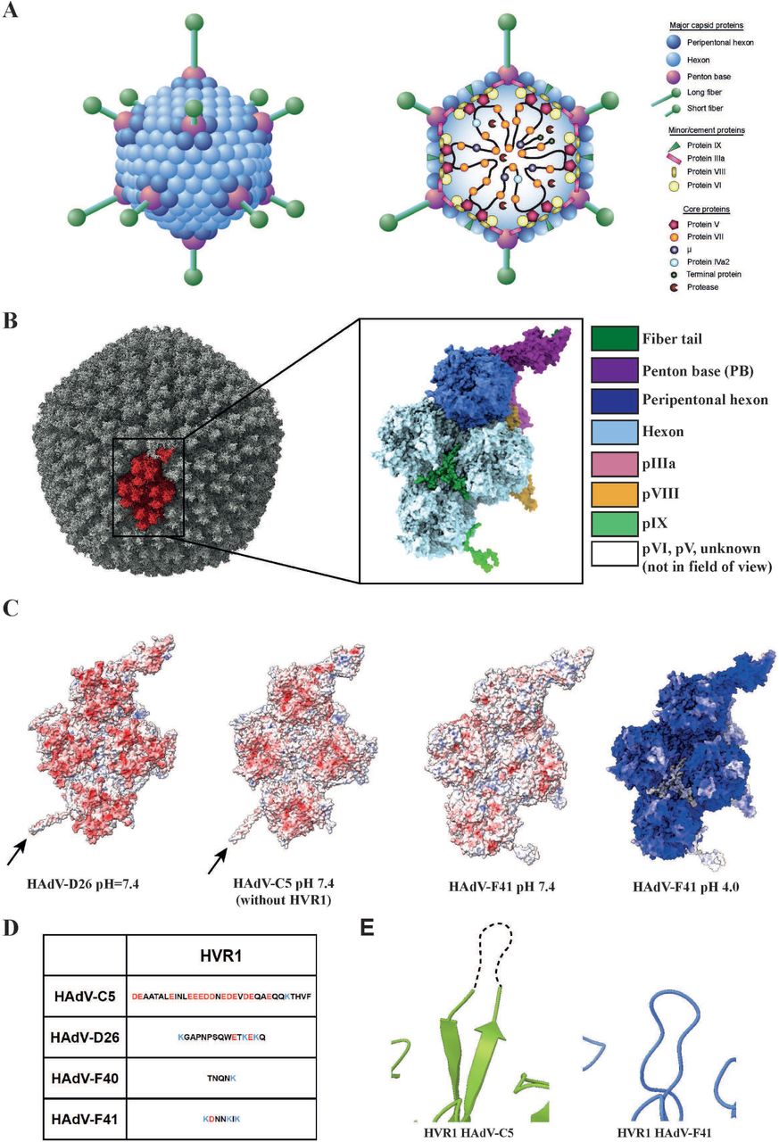
The enteric HAdVs have adapted to a distinct tissue tropism from other adenoviruses, which is presumably reflected in their capsid structure. One major known difference is that enteric HAdVs contain two different types of fibre proteins, long and short21,22, whereas other AdVs contain only one type of fibre.
Virions dock on to cells through fibre interactions with cellular receptors, followed by internalisation mediated by penton base interactions with cellular integrins23. All other HAdVs contain a conserved, integrin-interacting Arg-Gly-Asp (RGD) motif in the penton base24. Strikingly, the enteric HAdV-F40 and -F41 lack this conserved RGD motif and thus use different integrins for entry25, which may explain their different and much slower entry mechanism26,27.
Despite the medical importance of enteric adenoviruses as a major cause of childhood mortality through diarrhoea, the structural basis for their infection is not known. We used cryo-EM to determine near-atomic structures of the HAdV-F41 virion at pH=7.4 and at pH=4.0, the latter set as an average of the diurnal pH in the stomach of young children28.
These structures reveal the enteric adenovirus as having a pH-insensitive capsid with extensive surface remodelling as compared to non-enteric HAdVs. We further propose a conserved location of core protein V (pV), which links the adenovirus genome to the capsid. Lastly, we describe the assembly-induced structural changes to the penton base protein.
We believe that these findings will lay the foundation for a detailed understanding of enteric adenoviruses and how to prevent their infection.
reference link: https://www.biorxiv.org/content/10.1101/2020.07.01.181735v1.full
More information: K. Rafie et al. The structure of enteric human adenovirus 41—A leading cause of diarrhea in children, Science Advances (2021). DOI: 10.1126/sciadv.abe0974


