")


 Extract (6-MSITC) in Healthy Older Adults")
: An In-Depth Exploration into its Thermogenic Role and Social Significance")


Getting around without the need to concentrate on every step is something most of us can take for granted because our inner ears drive reflexes that make maintaining balance automatic.
However, for about 1.8 million adults worldwide with bilateral vestibular hypofunction (BVH) – loss of the inner ears’ sense of balance – walking requires constant attention to avoid a fall.
Now, Johns Hopkins Medicine researchers have shown that they can facilitate walking, relieve dizziness and improve quality of life in patients with BVH by surgically implanting a stimulator that electrically bypasses malfunctioning areas of the inner ear and partially restores the sensation of balance.
Results from their study of eight patients using the device are published today in the New England Journal of Medicine.
To maintain balance while moving through the world around us, our brains receive and process data from multiple sensory systems, including vision, proprioception (muscles and joints) and vestibular sensation from the inner ears.
People with BVH have difficulty keeping their eyes, head and body steady. Head movements make their vision jump and blur, and walking requires conscious effort. Forced to deal with this mental distraction, individuals with BVH suffer a more than thirtyfold increase in fall risk and the social stigma of appearing to walk like someone who’s intoxicated.
Current therapy for BVH is limited to vestibular rehabilitation exercises. Doctors advise their patients with BVH to avoid medications that damage the inner ear (ototoxic drugs) or suppress brain function (sedatives), and caution them to steer clear of activities that might endanger them or others, such as driving, swimming and walking in poorly lit areas.
“Although about 20 individuals had been implanted elsewhere with devices used to stimulate the vestibular nerve in a laboratory setting, participants in this trial are true pioneers – the first to use a vestibular implant as a long-term, 24-hour-per-day sensory restoration treatment,” says senior study author Charley Della Santina, M.D., Ph.D., professor of otolaryngology-head and neck surgery and biomedical engineering at the Johns Hopkins University School of Medicine and director of the Johns Hopkins Vestibular NeuroEngineering Laboratory, which conducted the study.
To achieve this milestone, Della Santina and his colleagues used basic research and engineering technology to modify a cochlear implant — a device that improves hearing loss by electrically stimulating the inner ear’s cochlear nerve — to instead activate the nearby vestibular nerve in response to signals from a motion sensor on the patient’s head.
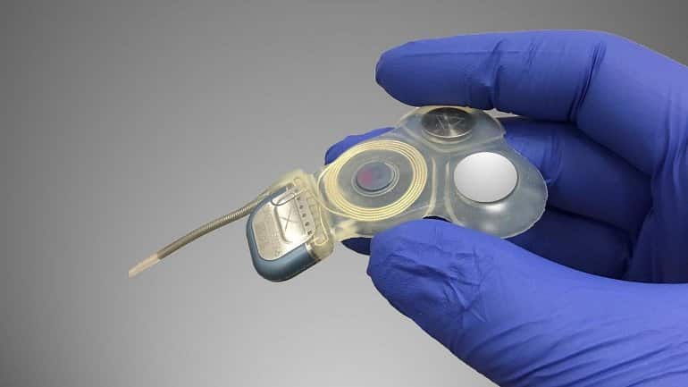
Electrical pulse strength and timing convey information about the speed and direction of the patient’s head motion which, in turn, drives head and eye reflexes that help maintain clearer vision during head movement and reduce the need to exert conscious effort to avoid falls.
In their study, the Johns Hopkins Medicine researchers evaluated eight patients with BVH who received the vestibular implant, assessing changes in postural stability, walking, hearing and patient-reported outcomes, including dizziness and quality of life.
Assessments were conducted before implantation surgery (the baseline measure) and at six months and one year afterward. Median scores improved for the group on four of the five posture and gait metrics, and on three of the four patient-reported outcomes.
All eight patients experienced some hearing loss in the implanted ear. Five maintained hearing in the implanted ear sufficient to use a telephone without a hearing aid, and three experienced greater hearing loss.
“Improvement in performance on standardized clinical tests of balance and walking has been remarkable,” says Margaret Chow, study lead author and biomedical engineering doctoral candidate at The Johns Hopkins University. “Even more gratifying is that our patients have been able to return to activities that enrich their daily lives, such as exercising, riding a bike, gardening or dancing at a daughter’s wedding.”
Overall, the improvement in quality of life and relief from the misery of BVH has been life altering, says A’ndrea Messer, Ph.D., one of the patients chronicled in the Johns Hopkins Medicine study and a senior science and research information officer at Penn State University.
“The multichannel vestibular implant is incredible,” says Messer. “Before receiving it, I couldn’t walk in the dark, on uneven ground or without a cane. Now, I can do all of those things and am living a fairly normal life.”
Along with Della Santina and Chow, the research team members from the Johns Hopkins University School of Medicine are Andrianna Ayiotis, Peter Boutros, Stephen Bowditch, John Carey, Yoav Gimmon, Carolina Treviño Guajardo, Kelly Lane, Brian Morris, Desi Schoo, Michael Schubert, Daniel Sun and Bryan Ward. Team members from industry sponsor Labyrinth Devices LLC are engineers Mehdi Rahman and Nicolas Valentin, both alumni of the Vestibular NeuroEngineering Laboratory.
The term bilateral vestibular loss refers to reduced or absent vestibular function on both sides due to deficits either in the labyrinths, in the vestibular nerves, or in a combination thereof. Because the deficit may be partial rather than complete, the term bilateral vestibular weakness is usually more appropriate, but the clinical picture is nevertheless usually designated bilateral vestibular loss in the literature.
Most patients with bilateral vestibular loss have either global reduction of all vestibular portions of the inner ear, such as gentamicin ototoxicity, or damage largely confined to the horizontal semicircular canals—bilateral vestibular neuritis.
We will use the term severe bilateral vestibular loss to denote a situation where between 75 and 100% of the inner ear is dead.
We will use the terms moderate for between 50 and 75% loss, and mild for 0 to 50% loss. In later sections of this article, we will discuss how one can estimate the degree of loss.
Clinical Features of Bilateral Vestibulopathy
Oscillopsia is the sensation that the environment moves when the head moves, but is still when the head is still. It is the clinical hallmark of bilateral vestibular loss because it is sensitive and specific. Oscillopsia is usually caused by loss of the normal vestibulo-ocular reflex (VOR). Oscillopsia is most noticeable with abrupt head movements, even when they are small (Fig. 1).
For instance, during normal ambulation there is a small degree of sway of the head due to the natural, rhythmic, alternating anterior-posterior flexion of the neck.
In a person with a normally functioning vestibular system, this movement of the head is exactly offset by equal and opposite movement of the eyes, so the person does not see any relative movement of the environment with respect to the head. In contrast, in an individual with bilateral vestibular loss, this small head movement is not offset by compensatory eye movements, so the person sees the environment sways up and down with each step. Other abrupt head movements are
similarly noticeable, such as when a person is riding in a car that goes over a bump.1
Imbalance
People with bilateral vestibular loss typically complain of imbalance. Imbalance is very common, and although it is required for diagnosis, it is not at all specific for bilateral loss. The reason for imbalance is straightforward: To “calculate” one’s position, velocity, and acceleration, the brain normally uses sensation from the inner ear, vision, and proprioception as well as its internal estimates based on motor efference.
In a person with bilateral vestibular loss, the brain attempts to compensate for the deficit by relying more heavily on internal estimates and remaining vision and proprioception. These may not always be as salient as the vestibular system, such as in the dark, or when one’s base of support is unsteady or narrow.
Therefore, persons with bilateral vestibular loss find it especially difficult to navigate through poorly illuminated environments, or walk on surfaces that are spongy. They are vulnerable to their other senses breaking down due to cataracts, neuropathy, or after insertion of joint prostheses. They are also more vulnerable to cognitive decline.

Hearing Loss
Surprisingly, hearing loss or tinnitus is not a regular accom- paniment of bilateral vestibular loss. This may be because the two main sources of bilateral loss—gentamicin ototoxicity and vestibular neuritis, generally have very little effect on hearing. The exceptions to this general rule are unusual conditions where the entire ear is damaged, such as from conditions that fill the inner ear with inflammatory cells (e.g., meningitis, Cogan’s syndrome).
Epidemiology and Etiologies
Fig. 2 shows data from our clinical practice in Chicago, Illinois. It illustrates that aminoglycoside ototoxicity has caused the giant share of bilateral loss. There have been several other series of patients with bilateral loss, reporting great variety in patient composition.2–4
Although we found about two-thirds of the patients in our practice had gentamicin ototoxicity, others report as few as 11%.2 We suspect that this difference is related to referral patterns. No study has yet been performed where a random sample of subjects were tested and diagnosed.

The diseases commonly cited as responsible for bilateral vestibular weakness include ototoxicity, meningitis, neurop- athy, bilateral vestibular disorders such as vestibular neuritis, autoimmune diseases, tumors, and idiopathic cases.5
Rinne and Bronstein reviewed 53 cases,3 and found 39% were associated with neurologic disorders (13% with cerebellar degeneration, 9% cranial or peripheral neuropathies, 11% with meningitis), 17% were attributed to gentamicin ototox- icity, 10% were associated with autoimmune disease, 6% were associated with malignant tumors, 8% were due to bilateral occurrence of otologic disease (Ménière’s disease, temporal bone fracture, Usher’s syndrome), and 21% were idiopathic. With respect to prevalence, again data are lacking.
In our otoneurological practice, over the past 20 years, we have identified ~300 patients out of 17,000 patients. In other words, roughly one in fifty patients in our otoneurology practice was ultimately diagnosed as having bilateral vestibular loss.
Familial bilateral vestibular loss, with or without hearing loss, was reviewed by Jen in a previous issue of this journal.6 Inherited bilateral vestibular loss is extremely rare.
Age In the clinic, bilateral loss is largely a disorder of older middle age, and in the group of patients shown in Fig. 2, the median age was 55. This age distribution is probably due to a combination of the age distribution of the general population combined with the likelihood of being exposed to the main cause of bilateral loss, gentamicin ototoxicity.
Additionally, the incidence of bilateral loss probably increases with age; as in older persons with ataxia, bilateral vestibular damage is frequent.7
The prevalence of balance problems increases with age.8–10 This may be in part due to the substantial age-related attrition of vestibular hair cells. Temporal bone histopatho- logical studies show that in otherwise healthy individuals there is a continuous decline of vestibular hair cells over the lifespan11 and 30 to 50% of both vestibular nerve fibers and vestibular hair cells are gone by the age of 80.12–14
A 30 to 50% vestibular deficit is generally well tolerated and is not a source of excess decline in otherwise healthy individuals,15 but when combined with another inner ear disease or with another sensory deficit such as visual or proprioceptive damage, it could be significant. In older persons with visual loss such as due to macular degeneration or cataract, the presenting symptom may be imbalance rather than oscillopsia.
Ototoxicity
Aminoglycoside Antibiotics
The aminoglycoside antibiotic, gentamicin, probably causes most cases of severe bilateral vestibular loss. Although all of the aminoglycoside antibiotics have ototoxic potential, some antibiotics, such as neomycin, are primarily cochleotoxic whereas others, such as gentamicin and streptomycin, are primarily vestibulotoxic.16
Of the latter, ototoxicity due to systemic gentamicin is probably the most frequent agent encountered in practices that see a high volume of patients with vestibular disorders, whereas other predominantly ves- tibulotoxic aminoglycosides (such as streptomycin) are rarely encountered.
Tobramycin is often the second most commonly encountered ototoxic antibiotic because of common use in cystic fibrosis. Fortunately, inhaled tobramycin is not associated with significant ototoxicity, and this problem is mainly confined to persons who have been given intravenous tobramycin.
The high prevalence of gentamicin ototoxicity, at least in the United States, is explained by several aspects of its pharmacology. First, gentamicin is a “silent” ototoxin in most individuals, as it does not produce telltale auditory warning signs of tinnitus or hearing loss. Second, although gentamicin is eliminated from the blood in hours, gentamicin accumulates in the inner ear over months.17
Because of the “integration” of gentamicin by the ear, toxicity can occur in persons in whom blood levels have always remained within normal limits. Third, gentamicin is toxic to the kidneys that eliminate it from the blood. Thus, gentamicin toxicity can cause a runaway situation where the kidneys are damaged, the drug level goes higher, and even more damage occurs.
Finally, the toxicity of gentamicin for the ear is potentiated by another commonly prescribed antibiotic, vancomycin. Addi- tionally, gentamicin is very inexpensive. Although gentamicin is toxic and complex to manage, nevertheless it may be life saving for many difficult and dangerous infections.
Several genes (NOS3, GSTZ1 and GSTP1) have been identified that confer particular susceptibility to gentamicin ototoxicity,18 but this information has no clinical use other than pointing out that gentamicin can have an idiosyncratic toxicity.
Although gentamicin ototoxicity generally occurs in the context of systemic administration, when a tympanic mem- brane perforation is present, topical ear drops, such as Cortisporin Otic® (DSM Pharmaceuticals, Inc., Greenville, NC), which contains neomycin, or Garamycin ophthalmic solution, used off-label can cause ototoxicity by passage through the middle ear and across the round window into the labyrinth.19–21
Chemotherapeutic Agents
Practically, although several chemotherapeutic agents are known to be cochleotoxic, chemotherapy is not a significant cause of vestibulotoxicity. In fact, only cisplatinum has well established vestibular toxicity.22 Vestibulotoxicity is rare with cisplatinum, perhaps because the other toxicities limit use.
Non-ototoxic Causes of Bilateral Vestibular Loss
Autoimmune Inner Ear Disease Unlike gentamicin ototoxicity, autoimmune inner ear disease (AIED) generally affects hearing and vestibular function in tandem. Autoimmune inner ear disease typically presents with subacute bilateral sensorineural hearing loss that is rapidly progressive (over weeks to months), but up to half of all patients also experience vestibular symptoms.23
The diagnosis is established when the hearing loss reverses or significantly improves after a course of steroids.24,25 There
are reports of cases that present only with vestibular symptoms and no hearing loss,26 but diagnosis is open to question
in such cases because there is no opportunity to assess for steroid-responsive hearing loss. Although several antibody based assays have been proposed for AIED, such as the HSP-70 heat-shock protein antibody, none has proved reliable.27
Treatment of AIED is immunosuppression or cochlear implantation.
Cogan syndrome’s28 is a subtype of AIED, with accompanying ocular manifestations. Cogan’s syndrome has a similar phenotype to postmeningitic hearing loss, as the labyrinth may be occluded with fibrous tissue. Another AIED subgroup includes patients who have had substantial inner ear surgery, as they can develop signs of damage including hearing loss and/or dizziness from the opposite side, attributed to a sympathetic autoimmune reaction similar to Vogt-Koyanagi-Harada syndrome of the eye.29 All AIED disorders are uncommon, but are not so incredibly rare that one can safely disregard their existence.
Meningitis
Meningitis damages hearing and vestibular function in tan- dem. Bilateral loss of vestibular function is generally encoun- tered only in persons with moderate or greater bilateral hearing loss. Whether the cause of a given case of meningitis is infectious, autoimmune or chemical,30 the inflammatory process can easily involve the labyrinth via the vestibular and cochlear aqueducts, resulting in deficits in vestibular func- tion.31
The vestibular and cochlear aqueducts are more patent in children than in adults, which may explain why meningitis more frequently causes inner ear deficits in children than in adults.32 In patients who have had bacterial meningitis, vestibular function is often gradually lost over years because the labyrinth becomes ossified.
The lack of fluid in the labyrinth may be seen on 1.5 or greater Tesla, T2 or construc- tive interference steady state magnetic resonance imaging (CISS MRI; see Fig. 3). One must often look at several sections to be sure that one is not missing seeing the labyrinth due to cut orientation.
Bilateral Vestibular Neuritis33,34
Vestibular neuritis typically affects primarily the superior vestibular nerve.35 Rarely, the inferior nerve is also involved,
as for example in the very severe vestibular neuritis that can accompany Ramsay Hunt syndrome. Nevertheless, the propensity of vestibular neuritis to spare the inferior vestibular nerve can be used for diagnosis as vestibular evoked myogenic potentials (VEMP) tests allow one to detect functional inferior vestibular nerves in persons with loss of caloric function and rotatory chair. This picture is the “diagnosis” in persons with complete or near complete loss of horizontal canal function and robust VEMP tests. Loss of calorics, by itself, is insufficiently specific: Caloric testing is very vulnerable to anatomical variables such as narrow ear canals, poor technique, and ear wax.
Bilateral Vestibular Schwannomas Bilateral vestibular loss can occur in neurofibromatosis type 2 where there are bilateral acoustic neuromas.36 This is extremely rare.
Bilateral Ménière’s Disease37
Ménière’s disease commonly results in bilateral hearing reduction that develops over decades,38 but bilateral pro-
found deafness is rare, and similarly, Ménière’s disease almost never causes the analog of profound deafness, profound bilateral vestibular loss. In Ménière’s disease, hearing is first affected, and labyrinthine function follows years later. Bilat- eral vestibular loss due to Ménière’s disease is not encountered in persons who retain “aidable” hearing.
Neurosyphilis
Otologic involvement in neurosyphilis generally presents with hearing loss, but in case series it is reported that from 4239 to 52%40 of patients also experience vertigo. Many of these cases appear to have bilateral vestibular weakness,40,41
with some case series reporting electronystagmographic abnormalities (typically vestibular weakness) in as high as 80% of patients.42
Neurosyphilis has almost disappeared due to increased use of antibiotics. For this, reason routine blood testing of patients with bilateral loss for syphilis is extremely low yield, but still considered reasonable as it is one of the few potentially treatable causes of bilateral loss.
Circulatory disturbances are extremely rare causes of bilateral vestibular loss, if indeed they ever occur at all. The inner ear blood supply is the labyrinthine artery, which is a branch of the anterior inferior cerebellar artery (AICA). For both labyrinths to become infarcted, one would need to experience ischemia of both labyrinthine arteries, an ex- tremely unlikely event. A selective loss of vestibular function on one side is very rare in AICA,43 and it would be nearly impossible for it to be bilateral.
Neurosarcoidosis
Neurosarcoidosis44 is an extremely rare cause of inner ear damage in general, and it has no particular predilection for the inner ear or the vestibular nerve. It is a very implausible source of bilateral vestibular loss. Blood testing of patients with bilateral loss for sarcoidosis, though commonly done, is almost never positive.
Congenital Malformations
Although a large number of congenital disorders are known to be associated with malformations of vestibular end organs,45
very few have been adequately studied to establish whether actual objective bilateral vestibular weakness is present.
The Mondini malformation and the CHARGE association (Coloboma of the eye, congenital Heart defects, choanal Atresia, mental and/or growth Retardation, Genital hypopla- sia, and Ear anomalies and/or deafness) are the most com-
monly associated with aplasia of the semicircular canals.46

These patients have congenital deafness and their computed tomography (CT) scan or MRI may show an absence of the labyrinth, similar to Fig. 3. Mondini malformations and related developmental abnormalities are common in persons
with bilateral congenital deafness.47
Head Trauma
Bilateral vestibular loss following head injury nearly always also includes hearing loss because the mechanism of injury is
labyrinthine concussion and/or a traction injury of the ves- tibulocochlear nerves,48 sometimes secondary to temporal bone fracture.49
The prevalence of traction injuries is unknown, but the “design” where the nerve exits through a bony canal and then almost immediately enters the brainstem could be vulnerable. The temporal bones are the hardest bone in the body, and for this reason, bilateral temporal bone fractures are generally also accompanied by brain injury.
Associations of Other Conditions with Bilateral Vestibular Loss
Migraine was associated with bilateral vestibulopathy in several case series.50,51 The main difficulty is distinguishing
ordinary migraine that occurs at a frequency of 14% in the general population from a putative “vestibulopathic” mi- graine.52 It has also been shown that migraine can be triggered by vertigo.53 At this writing, the situation is unclear.
Cerebellar Degeneration
There are several case series of cerebellar degeneration associated with bilateral vestibular weakness.3,54,55 As
both vestibular loss and cerebellar lesions cause ataxia, the vestibular portion of the disorder can be easily missed. When also accompanied by peripheral neuropathy, the syndrome may be labeled cerebellar ataxia, neuropathy, vestibular areflexia syndrome (CANVAS).54
In our clinical experience, these patients make up less than 1% of individuals with bilateral vestibular loss. Of course, prognosis for improve- ment with physical therapy is worse in patients with addi- tional neurologic deficits compared with pure bilateral vestibular loss.
Treatment for Bilateral Vestibular Loss
In the extremely rare situations where it is possible to affect the source of a bilateral vestibulopathy directly, such as stopping gentamicin, this is the first thing to do. On very rare occasions, one may identify a treatable cause of bilateral weakness, such as autoimmune inner ear disease.
Neverthless, it is unlikely that any medication will reverse or improve a bilateral loss. This is because hair cells and neurons of the human inner ear do not regenerate.68
Vestibular Rehabilitation Therapy
Nearly all patients with bilateral loss will improve with vestibular rehabilitation physical therapy.69 The therapy as- sists patients in compensating for their lost stream of vestibular sensory input.
One should avoid management of bilateral loss with vestibular suppressants, and avoid using other medications with vestibular suppression as side effects (antihistamines, tricyclic antidepressants, benzodiazepines).
These medications make the vestibular weakness worse. If an antidepres- sant is needed, one that is activating, such as venlafaxine, is preferred to one that is sedating such as amitriptyline. Benzodiazepines are useful for managing anxiety, but they should be avoided whenever possible as they suppress vestibular function, they may suppress compensation, and they increase fall risk.70
There is ongoing research on the development of vestibu- lar prostheses,71 but this treatment approach remains highly investigational in humans. A more promising approach is the regrowth of inner ear hair cells.72,73
Natural History of Bilateral Vestibular Loss
It is better to have less vestibular damage; of course, persons with mild bilateral loss do better than those with moderate or severe loss.74 In fact, persons with mild bilateral loss, here defined as having rotatory test results resembling persons with well-documented unilateral vestibular disorders such as vestibular nerve section—up to 50% loss—generally are indistinguishable from normal after 1 to 2 years of compen- sation, and have few or no complaints.
Persons who have moderate loss (50–75%) complain of oscillopsia and ataxia. Persons with severe bilateral loss (75–100%) have similar complaints, but are more limited in their activities of daily living.
For example, they often refuse to drive at night. Persons with bilateral loss who also have other impairments—such as a diabetic neuropathy, orthopedic disorder, visual disturbance, or cerebellar damage—do not recover their balance as well as those with an otherwise normal sensory and motor system.
People with bilateral vestibular loss usually improve, although the extent of improvement differs depending on the etiology.74,75 For those in whom bilateral vestibular weakness is the result of ototoxicity, after the first 3 months (damage may actually increase initially), there is measurable improvement in the vestibulo-ocular reflex up to roughly 2 years.76 Recovery is usually incomplete in cases that are moderate to severe.
More specifically, it is rare for persons with acute complete vestibular weakness, to recover to better than four lines on the DIE test. It is common for persons with any degree of bilateral weakness to return to driving, but rare for cases that are moderate to severe to return to driving at night. It is common for persons with bilateral weakness to return to work, if their work does not demand good balance.
Bilateral vestibular loss would ordi- narily prevent someone from returning to work to an occu- pation that requires good balance, such as a painter, electrician, or cable TV installer.
It is generally safe to advise newly diagnosed patients with ototoxicity, that they will get substantially better, but never return to normal. In idiopathic bilateral patients, it is frequently helpful to counsel them that even if their inner ear function is completely lost, from experience with similar patients with ototoxicity, most individuals, though limited in their balance, can continue to work at sedentary occupations.
Mechanisms for Improvement
It takes roughly 2 years to reach maximum medical im- provement. Possible mechanisms for improvement include central compensation, peripheral recovery, and behavioral adaptation.
Central compensation / plasticity means that the brain “listens harder” to remaining vestibular input, and increases its reliance on nonvestibular input to maintain balance.77
Animal data suggest that central compensation occurs pre-dominantly in brainstem vestibular nuclei,78 but functional
MRI (fMRI) evidence in humans suggests that there is also cortical reorganization.79
Recovery of vestibular hair cells can occur when there are hair cells that are damaged, but not dead. There is some evidence of this in animals,80 and it seems very likely that it also occurs in humans. Generally, any peripheral recovery is complete by 6 months.
Patients with bilateral loss learn behavioral adaptations. They avoid unsafe activities where their impaired balance or vision may result in danger. They do not stand on ladders or climb on cliffs. It is also very rare that patients with severe bilateral vestibular loss return to riding a bicycle or driving at night.
reference link: https://www.researchgate.net/publication/256931433
Original Research: Closed access.
“Posture, Gait, Quality of Life, and Hearing with a Vestibular Implant” by Charley Della Santina et al. NEJM


