


 Extract (6-MSITC) in Healthy Older Adults")
: An In-Depth Exploration into its Thermogenic Role and Social Significance")


The study, published today in the Journal of Cachexia, Sarcopenia and Muscle from King’s College London and the University of Southampton, is the first to identify differences in gut bacteria based on appetite between otherwise healthy older adults.
Researchers also found that lower appetite was associated with reduced muscle strength and function, with gut bacteria as a potential link between the two.
The team used appetite questionnaire answers to identify 102 older people who had poor appetite and 102 older people who had good appetite, and compared their gut bacteria. The two groups were otherwise as similar as possible in terms of age, body mass index, calorie consumption, antibiotic use and other factors that could impact gut bacteria.
The team then looked at participants’ muscle strength, based on previous muscle strength assessments completed during clinic visits, and found that twins with a poorer appetite had reduced muscle strength compared to twins with a good appetite.
Co-first author Dr Ruth Bowyer, Research Associate at TwinsUK, King’s College London, said: “Loss of appetite is very common in older people, and this can have serious consequences including loss of muscle mass and function. Our research is the first to explore the links between appetite and gut bacteria, and how this may be related to muscle strength.”
Co-first author Dr Natalie Cox, Clinical Research Fellow at the University of Southampton, explained: “A poor appetite can lead to poor nutrition and weight loss, which in turn can lead to loss of muscle bulk and so reduced muscle strength. We know from previous research however that a poor appetite is also linked to loss of muscle strength independent of overall weight loss.
“We now need studies to understand how exactly appetite, gut bacteria and muscle function affect each other and in what order. This could inform the development of treatments in the future to preserve muscle mass and function, to improve health in older age.”
Appetite regulation
The gastrointestinal (GI) tract is the body’s largest endocrine organ and the largest interface between the human body and the external environment(38). This external environment includes foods and the products of digestion, and the GI tract is thus one of the key players in the body’s regulation of appetite(39). The surface of the GI tract provides the means for detecting luminal nutrients and generates endocrine and neuronal signals to inform the body of their presence(40). Ultimately, these signals are transmitted to the central nervous system where they are integrated to orchestrate the short-term feelings of hunger and satiety.
Central regulation of appetite
The hypothalamus is regarded as the main ‘appetite centre’ in the central nervous system(41). Several hypothalamic regions have been shown to play a role in appetite regulation, but in particular, the arcuate nucleus (ARC) has been highlighted as important(42).
The ARC is strategically located near a region of the brain with an incomplete blood–brain barrier(43). This enables ARC to sense and integrate hormonal and metabolic signals from the peripheral circulation with the neuronal inputs from the central nervous system and periphery. The ARC contains two functionally opposing types of neurones involved in the regulation of energy balance: anorexigenic pro-opiomelanocortin (POMC) neurones and orexigenic neuropeptide Y (NPY)/agouti-related peptide (AgRP) neurones(44). Both types of neurones project to second-order neurones in other parts of hypothalamus and in extra-hypothalamic brain regions(42).
Following food intake, the POMC neurones are activated(45). Activation of these neurones indicates the ‘fed state’ and leads to a decrease in appetite and an increase in energy expenditure(44). Conversely, the ‘fasting state’ activates orexigenic NPY/AgRP neurones(46).
These neurons co-release NPY and AgRP which stimulate hunger and reduce energy expenditure(47). Acute pharmacological activation of NPY/AgRP neurones dramatically increases energy intake in mice(48). In addition, NPY/AgRP neurones directly inhibit the activity of POMC neurons in ARC through the release of inhibitory neurotransmitter γ-aminobutyric acid (Fig. 1)(49). The deletion of vesicular γ-aminobutyric acid transporter genes in the AgRP/NPY neurones results in a lean and obesity-resistant mouse, highlighting the physiological importance of this pathway(50).
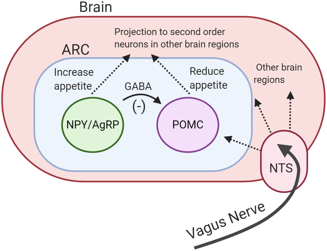
Neuronal signals from the GI tract are transmitted to the brain via the vagus nerve. The nucleus of the solitary tract in the brainstem receives vagal afferent signals and transmits these to downstream brain regions, including the hypothalamus(51). The importance of the vagus nerve has been highlighted by studies demonstrating that surgical transection of vagus nerve increases food intake and feeding duration in rodents(52, 53).
Peripheral control of appetite
Following food ingestion, signals are generated to increase the efficiency of digestion and reduce subsequent feeding and meal size. The entry of food in the GI tract (stomach and proximal small intestine) causes distention which stimulates mechanoreceptors(54). Activation of these receptors generates neuronal signals that act to slow gastric motility, allowing more time for digestion and creating a feeling of fullness(55). In addition to this, upper GI-hormones such as cholecystokinin are released in response to the presence of food. Together, these signals are thought to provide the initial, short-lived appetite suppression.
More sustained appetite suppression is believed to be brought about by the actions of other gut hormones. The two major anorexigenic hormones implicated in appetite regulation are peptide YY (PYY) and glucagon-like peptide-1 (GLP-1)(56). Both hormones are secreted from the intestinal enteroendocrine L-cells, which express the necessary molecular machinery to sense luminal nutrients and other digestive secretions such as bile acids(57).
The density of L-cells increases along the GI tract, with the highest numbers found in the colon. In response to the detection of nutrients and other digestive factors, L-cells release GLP-1 and PYY(58, 59). All three macronutrients, their by-products and other digestive factors such as bile acids have been shown to stimulate the release of both hormones(60).
PYY is a peptide hormone from the pancreatic polypeptide family. There are two biologically active forms of PYY in the human body(61). PYY (1-36) is released from L-cells and cleaved by dipeptidyl peptidase IV to form PYY (3-36). Anorectic effects of PYY (3-36) are believed to be mediated by the Y2 receptor, which is found throughout the central nervous system, including the ARC, as well as on vagal neurones(62, 63).
Binding of PYY to the Y2 receptors on NPY/AgRP neurones was shown to prevent their orexigenic activity and their inhibitory effects on POMC neurones, increasing anorexigenic activity(62). Increased levels of PYY decrease appetite, delay gastric emptying and reduce GI motility, contributing to the ‘ileal brake’(62, 64–66). Following a meal, a rise in PYY concentrations can be observed within 15 min.
Given that the L-cells are mainly located in the distal gut, this early phase release is believed to be the result of neuronal stimulation or perhaps another hormone acting on L-cells, rather than reflecting the effects of direct contact of nutrients with the L-cells(67). A second phase or peak of PYY is usually observed about 90 min following food intake, which is likely driven by the arrival of nutrients to the distal gut and their direct effects on L-cells(68).
GLP-1 is a peptide hormone that is also produced by intestinal L-cells, and by a population of neurones in the nucleus of the solitary tract of the brain stem. The nucleus of the solitary tract neurones are believed to be the primary source of GLP-1 in the brain, and have been shown to receive direct input from vagal afferents(69, 70).
The peripheral release of GLP-1 follows a similar biphasic pattern to PYY. Carbohydrate absorption in the proximal gut and other neuronal inputs are believed to contribute to its initial rise(71). GLP-1 acts as an incretin hormone through binding GLP-1 receptors on pancreatic β-cells to stimulate glucose-induced insulin release(72).
GLP-1 analogues such as Exenatide and Liraglutide have been approved as diabetes treatments since 2005 and 2010, respectively(73, 74). GLP-1 also slows gastric emptying, inhibits glucagon secretion and suppresses appetite(75, 76). GLP-1 receptors are found on the vagus nerve and in the brain including the ARC(77).
GLP-1 infusion in the hepatic portal vein has not been shown to change the activity of POMC or NPY/AgRP neurones, suggesting intestinal GLP-1 may not directly act on the ARC but rely on vagal afferent activation to modulate the central control of appetite(77, 78). The pathways by which GLP-1 and PYY work to modulate the central control of appetite are still unclear.
Obese individuals have been reported to have a blunted postprandial secretion or lower fasting levels of PYY and GLP-1(65, 79–82), which may contribute to weight gain. Bariatric surgery has also been shown to increase the postprandial release of PYY and GLP-1(83). This is believed to contribute to the dramatic weight loss observed following surgery(84).
As a result, peripheral administration of GLP-1 and/or PYY has been suggested as a way to correct the low levels observed in obese individuals to encourage weight reduction, or to mimic the weight loss effects of surgery. Indeed, peripheral administration of PYY and GLP-1 has been shown to suppress appetite and reduce food intake in both rodents and lean and obese human subjects(62, 64, 85, 86).
The chronic administration of these hormones also resulted in weight loss in animal and human models(62, 87–89). GLP-1 analogue Liraglutide has been approved as a weight loss treatment in patients without diabetes since 2015(90). These highlight that GLP-1 and PYY are key regulators of appetite regulation and that their manipulation may have an effect on energy intake and body weight.
Bacterial fermentation and energy metabolism
The human GI tract harbours a rich and dynamic community of microorganisms (gut microbiota) living in a symbiotic manner. Gut microbiota relies on hosts’ dietary intake as an energy source to grow and multiply. The main source of energy for microbiota is a dietary fibre which becomes available for bacterial fermentation in the distal gut. The complex interplay between dietary fibre, gut microbiota and microbial-produced metabolites has an impact on hosts’ energy metabolism. In relation to specific energy intake, these interactions can result in appetite suppression and reduced food intake, reducing the risk of excess energy intake and weight gain(30).
Dietary fibre and bacterial fermentation
Dietary fibre is an umbrella term for the group of carbohydrates that cannot be digested by the endogenous enzymes in the human body. There are several definitions of dietary fibre proposed by different countries and organisations(91). The most recent, and one of the most detailed, definitions has been made by the Australia New Zealand Food Authority(92): Dietary fibre is that fraction of the edible part of plants or their extracts, or synthetic analogues, that are resistant to digestion and absorption in the human small intestine, usually with complete or partial fermentation in the large intestine. Dietary fibre promotes one or more of these beneficial physiological effects: laxation, reduction in blood cholesterol, and/or modulation of blood glucose. The term includes polysaccharides, oligosaccharides (degrees of polymerization >2), and lignin.
Epidemiological studies have repeatedly identified that high-fibre diets are associated with lower body weight(24–27). This inverse relationship can also be observed for weight gain(93), visceral adiposity(94), cardiometabolic diseases(26), type 2 diabetes(95) and colorectal cancer(96). Clinical trials with animals and human subjects have investigated the causal role of fibre in these effects.
Animal studies find supplementation with dietary fibre reduces energy intake and protects against weight gain(97–100). A meta-analysis of twelve randomised controlled trials identified a reduction in body weight of obese/overweight human subjects when their diets were supplemented with soluble fibre(101).
Dietary fibre has been proposed to exert its benefits on host metabolism partly through its interaction with the gut microbiota. As previously stated, dietary fibres resist digestion in the human gut and reach the distal gut. The number of bacteria increases along the gut, with colon having the highest numbers.
Thus in the distal gut, bacteria is at a capacity to see marked bacterial fermentation of dietary fibre. This process yields energy for bacterial growth along with gases and the side products called SCFA. The most abundant SCFA are acetate (C2), propionate (C3) and butyrate (C4), which are present in the colon in the approximate ratio of 3:1:1 although this ratio depends on the amount and type of dietary fibre and the composition of gut microbiota(102).
While SCFA are waste by-products for the microbiota, for the host, their production represents the extraction of energy from the undigested material that would otherwise be wasted in the stool. Species that consume plant-rich, high-fibre diets such as gorillas (75–80 g/d) rely on this system as their main energy source(103).
In the western world human subjects, SCFA are estimated to contribute 2–10 % of daily energy intake(104). In the large intestine, most of the SCFA (mainly butyrate) are rapidly absorbed and used as an energy substrate by the colonocytes. The remaining SCFA reach the liver via hepatic portal vein where they are used as substrates for gluconeogenesis or metabolised through other pathways(105). Only a small proportion of SCFA enter the peripheral circulation. A study quantified systemic availability of colonic acetate, propionate and butyrate as 36, 9 and 2 %, respectively(106).
SCFA and appetite regulation
In the human body, SCFA are more than merely a source of energy. SCFA have been shown to act as signalling molecules through their interactions with the NEFA receptors 2 and 3 (FFAR2 and FFAR3). Acetate and propionate activate FFAR2, whereas FFAR3 can also be activated by butyrate(107–109). FFAR are expressed in key areas involved in the regulation of energy metabolism, including the gut, adipose tissue and skeletal muscle(107, 110, 111).
In the gut, the FFAR are expressed in GLP-1 and PYY secreting L-cells. This discovery led to the suggestion that SCFA could stimulate the release of appetite-suppressing hormones which could explain the mechanism by which fibre influences body weight. Indeed, studies using rodent and human cell lines confirmed SCFA acted on FFAR on L-cells to stimulate the release of GLP-1 and PYY(30, 112, 113).
This finding was further strengthened by FFAR2 and/or FFAR3 knock-out rodent models which showed attenuated GLP-1 and PYY secretions in response to intra-colonic propionate infusions(112, 114, 115). Dietary supplementation with fermentable carbohydrates or SCFA increased the circulating concentrations of PYY and GLP-1 in animal models and activated neurones in hypothalamic regions involved in appetite regulation(98, 114, 116–118).
SCFA and energy intake
Despite the well-established link between SCFA and anorectic gut hormone release, the effect of SCFA on energy intake has been inconsistent. Several studies investigated the effect of supplementing rodent diets with SCFA on energy intake and found no effect(119–122).
One study supplemented high-fat diets with acetate, butyrate or propionate and showed a significant reduction in energy intake of mice following butyrate and propionate supplementations (22 and 9 %, respectively)(115). However, studies using oral supplementation of SCFA should be interpreted with care as the bitter taste of these supplements may cause food aversion. In another study, intragastric gavage of butyrate was found to significantly reduce food intake of mice(123). Frost et al. also demonstrated that intraperitoneal administration of acetate acutely reduced food intake for 2 h although colonic administrations showed no effect(124).
Oral SCFA failed to show an effect on food intake in human subjects(125). This may be due to SCFA being quickly absorbed in the upper gut, and thus not reaching the distal gut where they are proposed to interact with the L-cells to drive anorectic hormone release. Our research group has used inulin-propionate ester for the targeted delivery of propionate to the distal gut.
In a randomised controlled study, 7 d supplementation with the inulin-propionate resulted in a reduction in ad libitum energy intake(126). In another study, consumption of 10 g inulin-propionate ester reduced ad libitum food intake and increased plasma GLP-1 and PYY concentrations compared to an inulin control(30). These results suggest that not oral but targeted colonic administration of SCFA may reduce food consumption in human subjects.
SCFA and body weight
Animal studies indicated a beneficial effect of SCFA on body weight. Studies with rodents highlighted that colonic infusions, intragastric gavage and oral supplementation of SCFA attenuated high-fat diet-induced weight gain(105, 120–123, 127–129). Lu et al. supplemented high-fat diets with acetate, propionate, butyrate or a mixture of all three SCFA, and fed these to mice for 16 weeks.
All SCFA significantly attenuated high-fat diet-induced weight gain, with acetate having the largest effect (72 % less weight gain)(128). In one study, germ-free mice received faecal transplants from obese and lean human subjects which resulted in the development of a similar phenotype in the recipient mice(130).
The lean mice had significantly higher caecal levels of propionate and butyrate, suggesting a potential benefit of SCFA in mediating the observed difference in body weight(130). Similar results were observed following faecal transplantation between mice that undergone Roux-en-Y gastric bypass and germ-free mice(131). However, other studies have found weight gain following acetate supplementation(132, 133). In addition, studies using FFAR2/3 knock out animal models reported inconsistent outcomes on body weight(115, 134).
Only one human study has investigated the link with SCFA and body weight. Long-term (24 weeks) supplementation of inulin-propionate ester was shown to significantly reduce body weight gain in overweight participants compared to the inulin control as part of a habitual diet(30). Only 4 % of participants in the inulin-propionate group gained significant weight (>3 % body weight) compared to 25 % in the control group.
Overall, these data highlight that SCFA stimulate the release of appetite-suppressing hormones. In human subjects, increasing colonic levels of SCFA may reduce food intake and protect against weight gain. Manipulating the colonic levels of SCFA is also possible through dietary modification since increasing fibre consumption has been shown to increase SCFA production. However, in addition to the fibre content, gut microbial composition can have a strong impact on SCFA production.
Gut microbiota composition and its effect on fermentation
Gut microbiota is a combined community of several types of microorganisms, including viruses, yeast and bacteria, with the latter being the most heavily researched and the most abundant(135). This is why the gut microbiota is sometimes referred to as the gut bacteria.
The human gut microbiota is dominated by six bacterial phyla: Bacteroidetes, Firmicutes, Proteobacteria, Verrucomicrobia, Fusobacteria and Actinobacteria, with the Bacteroidetes and Firmicutes making up about 90 % of the whole community(136, 137). Although the complete repertoire of the gut microbiota remains unrevealed, more than 500 species are estimated to reside in the GI tract(138).
The composition of the gut microbiota dictates its overall metabolism and functional capabilities including SCFA production. Indeed, the gut microbial composition has been shown to lead variations in SCFA productions(21, 139–142). However, it should be noted that this area of research mainly relies on in vitro models or stool SCFA measurements due to a lack of studies directly measuring luminal SCFA production in healthy human subjects.
Many bacterial species are capable of producing acetate whereas propionate and butyrate productions are more conserved and substrate-dependent. Butyrate producers are mainly from the Firmicutes phylum(137, 143, 144). Faecalibacterium prausnitzii, Eubacterium rectale and Eubacterium hallii are primary butyrate-producing species in the human gut(143, 144).
Resistant starch fermentation is believed to contribute to butyrate production by generating intermediate products that are fermentable by butyrate producers. Ruminococcus bromii (Firmicutes) and Bifidobacterium (Actinobacteria) are regarded as the main resistant starch-degrading bacteria(145–147).
Individuals with higher R. bromii abundance have been shown to produce more total SCFA and butyrate(140, 142, 146). Similarly, it has been shown that individuals with a higher Firmicutes abundance have a greater SCFA production capacity marked by increased acetate and butyrate productions(21, 139, 142, 148).
Although shared by a number of phyla, Bacteroidetes dominates the propionate producers(137, 144, 149). In line with this, one study showed a positive correlation between faecal propionate concentrations and the abundance of Bacteroidetes (141). Other species such as Akkermansia muciniphila and the phylum Firmicutes (mainly class Negativicutes) have also been associated with propionate production(149, 150).
In particular, A. muciniphila has been identified as a key propionate producer and drew attention as a potential probiotic due to its negative correlations with diabetes and obesity(150–153). Bacteroidetes mainly produce propionate from the fermentation of polysaccharides although they are also able to ferment peptides(144). Accordingly, diets high in protein and lower in fibre have been associated with an increase in Bacteroidetes, reflecting a switch from carbohydrate to protein fermentation(154, 155).
Although relatively stable during adulthood, gut microbiota is susceptible to changes by dietary intake, which can in turn affect SCFA production. In diet switching studies, it was elegantly shown that the composition of the gut microbiota and the magnitude of bacterial fermentation can be altered using high- and low-fibre diets(29, 155, 156).
High-fibre diets increase SCFA production and increase gut microbiota diversity(155, 157–161). Unsurprisingly these diets also stimulate the growth of carbohydrate fermenting bacterial species from the Firmicutes phylum(155, 158, 162, 163). Diets high in resistant starch were found to stimulate the growth of R. bromii and Bifidobacterium (141, 157, 159, 162–165). Conversely, western-style low-fibre, high-protein/fat diets lowered bacterial fermentation, reduced bacterial diversity and increased the numbers of Bacteroidetes (154, 155, 161, 166).
These studies highlight that diet and gut microbiota act together to impact SCFA production, and thus the subsequent effects of SCFA in the human body, including energy intake and body weight. Differences have been observed between the gut microbial composition of lean and obese subjects, highlighting the potential impact of gut microbiota on body weight(167–169). In addition, bariatric surgery has been shown to modulate gut microbial composition which is proposed to contribute to the weight loss following surgery(170, 171). However, this topic is beyond the scope of the present paper and reviewed in detail elsewhere(172, 173).
Food structure
Nutritional research has largely considered the effect of food and diets on human health based on the chemical compositions of foods (i.e. macronutrients and energy). However, this approach alone is insufficient as demonstrated by the variability in metabolic response to foods with the same energy and macronutrient profiles. Beyond macronutrient composition, food structure is fundamental for dictating the food behaviour, and thus how it is digested and processed within the GI tract. This in turn can impact on postprandial response and metabolism, such as microbial fermentation and appetite control.
Food structure and digestion
Food structure relates to the assembly of molecules making up food which can be a result of natural formation, domestic processing (cooking, blending, etc.), industrial processing or a combination. There are different levels of food structures which can impact digestion and subsequent postprandial metabolism. As previously mentioned, the main substrate for microbial fermentation is carbohydrate. Therefore, this review will focus on carbohydrate structures.
Molecular level: starch
Starch is a glucose polymer, and the main form of carbohydrate storage within plants. The glucose units can be linked with α-1,4 glycosidic bonds to form straight helical chains called amylose(174). Alternatively, the glucose units can be linked with a combination of α-1,4 and α-1,6 glycosidic bonds to form branched polymers called amylopectin. The proportion of these starch configurations differs between different plants and starch types(175).
In general, starchy foods such as barley, rice, wheat would contain 20–30 % amylose and 70–80 % amylopectin(176). Amylose is a straight chain, and thus with less surface area available for enzymatic actions, its digestion is slower compared to the highly branched amylopectin(177).
In vitro digestion studies have demonstrated this eloquently, for example, the digestion rate of different rice grains being shown to increase with the increasing ratio of amylose to amylopectin(178). In clinical human studies, high amylose starch supplementation resulted in attenuated postprandial glucose and insulin responses compared to high amylopectin starch, indicative of slower digestion from the former(179, 180).
At greater length scale, amylose and amylopectin chains are further assembled into granules consisting of a ratio of highly organised, dense pseudo-crystalline regions and less organised amorphous structures(181). The pseudo-crystalline regions are abundant in raw vegetables such as potatoes and unripe bananas and they are more resistant to digestion by α-amylase resulting in slower and sometimes incomplete digestion. In a study that investigated the in vitro digestion of potato, pea, maize, rice, barley and wheat starches, the digestion rate was found to decrease with the increasing amount of crystalline structures(182).
Food cellular structure
At its most basic, food cellular structures include animal, fungal and plant cells. Unlike animal cells, plant cells have cell walls providing structural support and shielding intracellular nutrients(183). These cell walls are made up of indigestible carbohydrates (i.e. fibres) such as cellulose, hemicellulose, pectin and non-carbohydrates such as lignin, proteins and water(184).
The relative proportion of these building blocks differs based on the type, function and maturity of plant tissue, which in turn dictates the permeability and strength of the cell wall and the digestive fate of these foods(185). For example, high amounts of cell wall lignin relate to the string-like structure of asparagus reducing the permeability of cell wall and thus hindering digestive enzyme access and nutrient bioaccessibility(186).
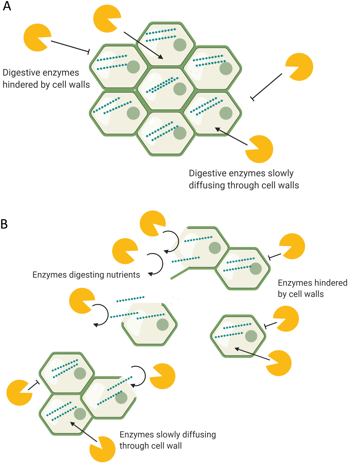
Bioaccessibility is defined as the proportion of consumed nutrients available for absorption in the human gut. Food cellular structures have been shown to impact bioaccessibility(187). Digestive enzymes need direct contact with the nutrients inside the cellular structures to be able to digest them. Endogenous enzymes in the human body are unable to digest plant cell walls (i.e. fibre), and therefore enzymes can only act to break down macronutrients within plant cells if they diffuse through the cell walls or if the cell walls are ruptured as part of the digestion process.
As a result, plants with strong and less permeable cell walls may undergo more attenuated and incomplete digestion within the GI tract which results in a slower and lower nutrient release. In vitro digestion studies have shown slower hydrolysis of cellular starch compared to extracellular starch(31), and in applied food examples, kidney bean and chickpea cells, which have remained intact following cooking, have resulted in a slower and incomplete in vitro digestion(181).
In vivo, human studies demonstrated an attenuated postprandial glycaemia and lipidaemia following the consumption of foods with intact cellular structures compared to macronutrient and energy-matched foods with disrupted structures, indicating a slower nutrient release from the former(31, 188–190).
In some cases, intact cells may protect cellular starch from digestion both of which reach the distal gut. Ileal samples from healthy human subjects were found to contain intact cells with cellular starch molecules, indicating that these cells escaped digestion in the upper gut(191). Food cellular structures are further organised into tissues at greater length scales, which further dictates their digestive behaviour.
Mechanical processing in human body
During digestion, food structures are altered due to mechanical stress, actions of digestive enzymes and physicochemical conditions of the GI tract such as pH(192). Oral processing (chewing) is the first stage of digestion and reduces the particle size of food, thus increasing the surface area for enzymatic digestion. This process changes the food matrices by separating tissues, and on a cellular level separating and/or rupturing cell wall surfaces.
Depending on cell wall structure and conformation, such structures have different strengths of intracellular adherence and tendencies to rupture or separate under mechanical stress(193). For example, high pectin levels in the cell walls usually indicate greater potential for cell separation. Under digestion, the cells of nuts, raw hard vegetables and seeds have a greater tendency to rupture, whereas cells of cooked foods such as legumes tend to separate and remain intact(194, 195).
This means that most legume cells remain intact following chewing, the result of which can provide one possible mechanism to explain their attenuated postprandial glycaemic response, due to lower enzymatic exposure to intracellular starch. Conversely, ruptured cell walls provide greater enzymatic exposure to intracellular starch (Fig. 2). Studies where foods have been swallowed without oral processing have demonstrated such, through reduced postprandial glycaemia, indicating a slower nutrient release due to the protection of cell walls(196).
Mechanical digestion continues in the stomach and small intestine where the digesta is constantly mixed by peristaltic gut movements. Plant foods with strong food structures can endure the contractions created by the GI tract and reach the distal gut relatively undigested.
One study using intubation technique to collect samples from the distal ileum of healthy volunteers found ileal digesta contained intact bean cells encapsulating starch molecules, indicating that these cells were resistant to digestion in the upper GI tract(191). Another study with healthy ileostomy patients demonstrated intact carrot cells containing intracellular nutrients in the ileal effluents(197).
Such findings, further highlighting intact cellular structures with encapsulated nutrients reaching the distal ileum, have been repeatedly confirmed within ileostomy patients(31, 198–200). Theoretically, the greater the protective effect of food structure, the greater is the amount of fermentable substrates delivered to the distal gut for fermentation. This may be indicating the beneficial role of food structure in stimulating appetite suppression through the proposed mechanisms within this review.
Industrial and domestic processing
Food processing has been shown to change food structures, typically leading to more digestible products. Thermal treatment in the presence of water (boiling) has been shown to cause gelatinisation of starch(201). Gelatinisation results in the loss of intermolecular bonds and pseudo crystalline structures, resulting in a more digestible compound. For example, raw potato starch or raw oats eaten as muesli are almost completely indigestible to human subjects.
Upon cooking, the starch in these foods gelatinises and can be easily digested, marked by a higher postprandial glycaemia(181). However, if the gelatinised starch is left to cool down, the dissociated starch molecules randomly re-crystallise in a disorganised manner (retrograding). This generates compounds more resistant to digestion (resistant starch). In a study with healthy ileostomy patients, cooked potatoes resulted in 3 % starch losses in the ileal effluents whereas cooked and cooled potatoes resulted in 12 %(202).
Beyond starch structures, processing has been shown to alter cell walls, typically leading to more permeable, weak or ruptured structures(31–37). For example, the fine milling process has been shown to rupture cell walls(31, 203). Others such as homogenisation, canning and cooking were shown to denature cell walls leading to rupture or weakening(185).
As previously explained, without the hindrance of rigid, impermeable cell walls, digestive enzymes can easily access the intracellular nutrients and hydrolyse them into absorbable molecules. Weak cell walls may also be more susceptible to rupture under mechanical digestion in the GI tract.
This can lead to more digestible and bioaccessible food products(187). Singh and coworkers reviewed the effect of different types of food processing on starch digestibility and found an increase in digestibility with processing(115). In another study, in vitro digestion of whole and finely milled almonds resulted in higher energy extraction from the milled almonds(198).
This was supported by microscopic analysis of almonds finding ruptured almond cells following the milling process while the whole almonds contained intact cells with encapsulated nutrients. Another in vitro digestion study estimated that only 5⋅5 % of lipids were released from whole almonds compared to 42 % from almond starch(204).
These results were repeated in a healthy ileostomy patient who digested 96⋅5 % of lipids from milled almonds compared to only 56⋅5 % from whole almonds(37). Increased nutrient availability of processed foods has been demonstrated by other studies using cooked, pasteurised and milled products(31–36).
More efficient digestion and absorption in the upper gut translates into lower levels of nutrients reaching the distal gut. In a study with healthy ileostomy patients, Livesey et al. demonstrated that the starch losses in the ileal effluents decreased 3-fold following the consumption of finely milled barley compared to coarse barley(199). An analysis of the ileal effluents found that the lost nutrients were still encapsulated within the intact cellular structures.
Langkilde et al. repeated this finding in an experiment where ileostomy patients were fed cooked and raw banana starch(36). Raw banana starch consumption resulted in more than three times higher amounts of starch losses in the ileal effluents (6⋅3 (sd 0⋅4) v. 21⋅4 (sd 0⋅6)).
Intact cellular structures with encapsulated nutrients were found in the ileal effluents of raw banana starch group. This indicated that the observed differences in both of these studies could be due to more nutrients being absorbed from the disrupted structures of processed foods while the intact cellular structures protected the nutrients in the raw diets.
Two studies showed contradictory results. Edwards et al. fed coarse and finely milled porridge to healthy ileostomy patients and found no difference in the starch losses in the ileal effluents(31). This was explained by the finding of intact but empty cellular structures in the effluents following the consumption of coarse oats.
Combined with the in vitro findings, this indicated that the cell walls were permeable to digestive enzymes although they were resistant to digestion. Another study found no difference between the ileal effluents following cooked and raw carrot consumption(197). This was explained by the failure of cooking to disrupt cellular structures and highlighted that not all food processing results in the disruption of cellular structures.
Food structure, microbial fermentation and appetite control
It is well established that food structures dictate the digestive fate of consumed nutrients(192). It is therefore unsurprising that food structures can also influence appetite control. Foods with rigid structures may require a longer chewing time while others may be consumed rapidly. Accordingly, diets high in plant matter (fibre) have been shown to reduce eating rate(144).
A slow eating rate prolongs the oral exposure of food which transmits orosensory satiety signals to the brain, resulting in a prolonged appetite suppression(145). The importance of this is demonstrated by the attenuated appetite suppression observed following direct infusion of foods into the stomach or duodenum(146, 147).
Li et al. also demonstrated that increasing oral exposure of food (15 v. 40 chews) resulted in higher postprandial GLP-1 concentrations and reduced ad libitum energy intake in obese and lean subjects(148). In another study, eating at a slower rate increased the postprandial GLP-1 and PYY levels(149).
Food structure was also found to impact gastric emptying which contributes to appetite suppression. In a randomised cross-over study, Mackie and coworkers demonstrated slower gastric emptying following the consumption of semi-solid meals compared to liquid(150).
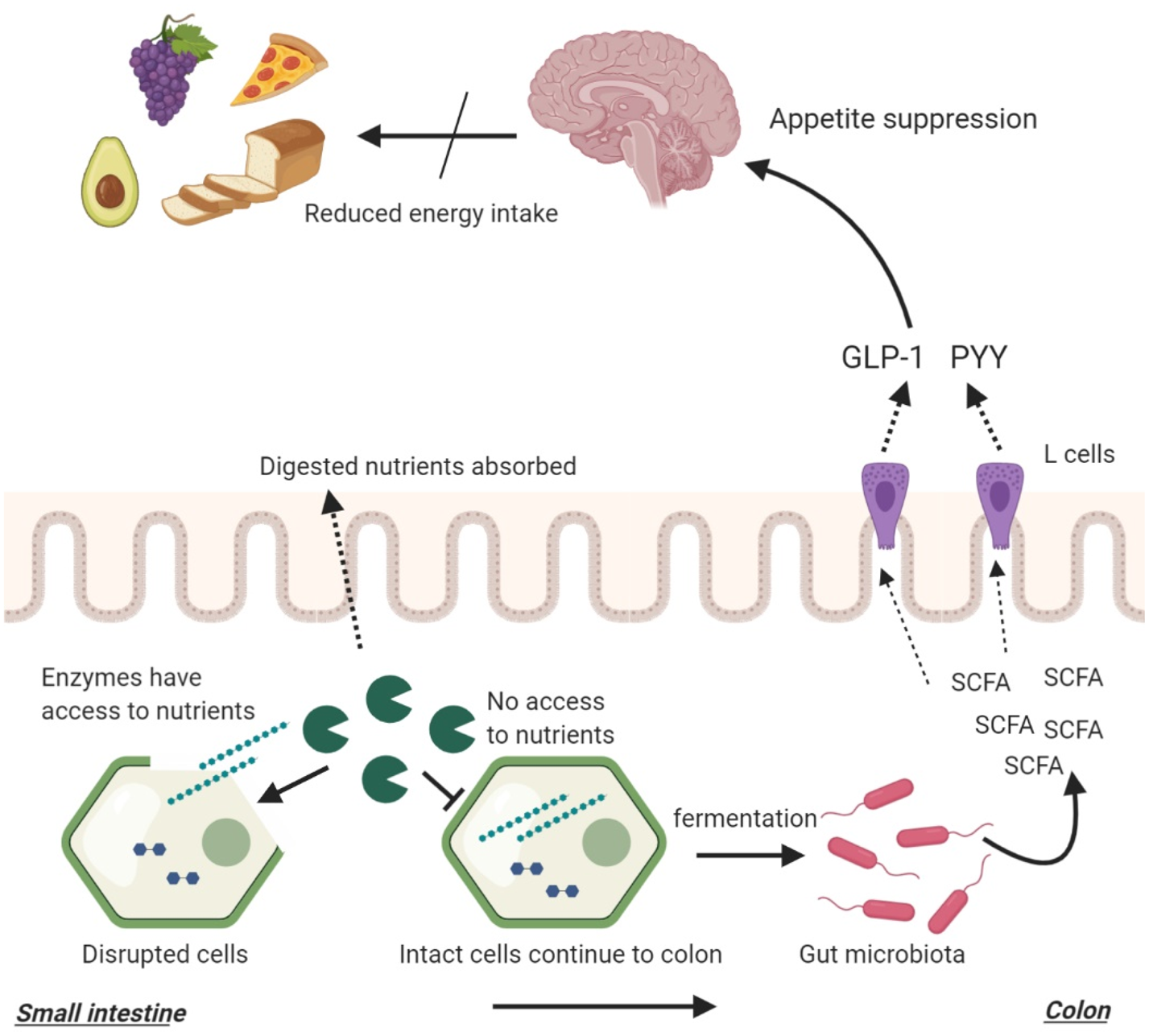
As previously described, food structures can alter the amount of nutrients reaching the distal gut which can impact bacterial fermentation (Fig. 3). While cell walls are indigestible by human enzymes, they are susceptible to bacterial enzymes and fermentation, producing SCFA(162). The composition of the plant cell wall is a major determinant of bacterial fermentation since different fibres have different fermentation capacities.
Complex, insoluble fibres such as cellulose and hemicellulose are fermented slowly and poorly by the gut bacteria whereas soluble fibres such as pectin and β-glucan are highly fermentable(162, 205). Bacterial degradation of plant cell walls may also expose the intracellular starch that also acts as an efficient substrate for bacterial fermentation. However, as previously mentioned, gut microbiota composition is also a determinant of SCFA production. The presence of certain microbial groups may enhance the fermentation of complex structures such as resistant starches(145–147).
In one study, ileal effluents with higher fibre and starch losses were found to result in higher in vitro SCFA production(206). This study highlights that the amount of nutrients reaching the distal gut is a determinant of microbial fermentation. In a previously mentioned study, ileal effluents were collected from healthy ileostomy patients following the consumption of raw or cooked banana starch(36).
The in vitro fermentation of the effluents resulted in significantly higher SCFA production following the inoculation of raw banana effluents which contained higher amounts of fibre and starch. In another study, the effect of food processing was investigated using native and finely milled whole grains(207). It was found that processing reduced the resistant starch components of foods which in turn reduced their in vitro SCFA production.
These studies highlighted that macronutrient, fibre and energy-matched foods can lead to different microbial fermentation due to a difference in food structure brought by food processing with more ‘resistant’ food structures resulting in greater SCFA production (Fig. 3). In addition, it highlighted that cooking and milling (processing) can reduce the amount of fibre/starch reaching the distal gut and reduce bacterial fermentation through increasing digestibility. This can in return reduce appetite suppression.
These findings indicate that generally, processed foods could be less satiating compared to raw, unprocessed alternatives. Evidence from cross-sectional studies suggests that high consumption of highly processed foods is associated with excess body weight(208–213). One study found an inverse correlation between the degree of processing and the satiety index of ninety-eight commonly consumed foods(214).
Another study used subjective measures of appetite and demonstrated higher satiety following the consumption of raw carrots compared to cooked carrots(215). In a randomised cross-over trial, Mori et al. demonstrated that whole almonds reduced ad libitum food intake in healthy volunteers more than energy, macronutrient and fibre-matched almond butter(216). The authors commented that this difference may be due to increased orosensory time and reduced gastric emptying.
In another study, it was shown that supplementation with native banana starch, which has been previously shown to carry intact cellular structures to the distal gut(36), reduced ad libitum food intake compared to supplementation with available banana starch indicative of appetite suppression(217). In a recently published randomised cross-over study, Hall et al. fed participants energy, macronutrient and fibre-matched ultra-processed (as per NOVA classification) and unprocessed diets for 14 d(218).
Ad libitum energy intake was found to be about 2092 kJ (500 kcal)/d greater in the ultra-processed diet group. This translated into participants’ body weight and fat mass increase in the ultra-processed diet group. Fasting PYY levels were also found to be significantly higher in the unprocessed diet group. Furthermore, the eating rate was also significantly lower in this group.
However, evidence in this area remains inconclusive and limited. There are still no studies investigating the causal link between food structures, bacterial fermentation and appetite suppression directly, with current hypothesis being based on the mechanistic link proposed in this review.
reference link: https://www.cambridge.org/core/journals/proceedings-of-the-nutrition-society/article/understanding-the-interplay-between-food-structure-intestinal-bacterial-fermentation-and-appetite-control/7A854AAFAF3B50A2C5FA273157DE4A6D
Source: King’s College London


