


 Extract (6-MSITC) in Healthy Older Adults")
: An In-Depth Exploration into its Thermogenic Role and Social Significance")


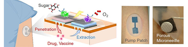
Researchers from Tohoku University have developed a biobattery-powered device capable of both delivering large molecule pharmaceuticals across the skin barrier and extracting interstitial fluid for diagnostic purposes.
They published their approach online on Jan. 28 in Nature Communications.
The team, led by corresponding author Matsuhiko Nishizawa, professor in the Department of Finemechanics in the Graduate School of Engineering at Tohoku University, developed a microneedle array smaller than a pinky nail.
The microneedles are porous, acting as interconnected conduits to either inject or extract fluid, including the large molecules of vaccines or even insulin.
That’s a step further than the patches already commercialized for small-molecule drug transmission used for post-operative pain relief or migraine treatments, Nishizawa said, without increasing needle size.
The porous microneedles are 250 microns long, about the width of three and a half human hairs—deep enough to painlessly penetrate the outermost layer of skin without being long enough to hit blood vessels or nerves.
When a low-grade voltage is applied to the array of porous microneedles coated with hydrogel, the flow of liquid is generated like when a syringe plunger is deployed.
Called electroosmotic flow, it can increase the transmission rate of drugs across the skin barrier or the extraction speed of interstitial fluid to be tested for such things as glucose levels.
The researchers powered the array with a biobattery. The battery consists of four coupled electrodes and converts chemical energy, taken from enzymes processing sugar and atmospheric oxygen, into electrical energy. The biobattery and porous microneedle array are secured to the skin with an adhesive patch smaller than a business card.
The researchers are now developing a porous microneedle array with a biodegradable polymer.
“The successful demonstration shown here using a built-in biobattery proves the future possibility of a totally organic electroosmosis flow-based skin patch that is safe and truly disposable,” Nishizawa said.
Telemedicine with home healthcare systems has attracted attention as an essential technology for addressing the growing problems of an aging society and for improving medical care during disasters/infectious epidemics1,2. Skin patches for transdermal diagnosis/treatment are typical devices for home healthcare, and there has been progress in improving skin compatibility, multifunctionality, and disposability3,4,5.
Iontophoresis has been studied to accelerate transdermal penetration/extraction by typically applying a continuous low voltage current6,7. The iontophoretic transdermal drug delivery systems have been actively studied8,9,10,11,12,13, and are already commercialized for the fast dosing of drugs for dermal anesthesia (LidoSiteTM, Vyteris Inc.)8, post-operative pain relief (IonsysTM, Alza)9, and anti-migraine (ZecuityTM, NuPathe Inc.)10.
Recently, a totally organic transdermal drug delivery patch containing a built-in enzymatic battery has been reported11. Also, the transdermal iontophoretic extraction of interstitial fluid (ISF) (a process sometimes called ‘reverse iontophoresis’) has been gaining attention as a sample collection method for medical diagnosis14,15,16,17, including continuous glucose monitoring with the GlucoWatchⓇ system18.
The mechanism of iontophoretic transport consists of the electroosmotic flow (EOF) of the solvent (water) as well as the electrophoresis of the charged molecules themselves. EOF is generated by the preferential movement of mobile cations (or anions) in the fluid conduits containing fixed anions (or cations) (Supplementary Fig. 1). Under physiological conditions, the skin acts as a cation-selective matrix (isoelectric point: ~4.5)19, and therefore an EOF can be generated in the anode-to-cathode direction (i.e., in the same direction as migration of cations)6,7.
The technical difficulty of these transdermal iontophoresis applications arises from the barrier functions of the stratum corneum, the outermost layer of skin (~20 μm thickness)7,17. Since iontophoresis is a DC technique, the electrical barrier of the stratum corneum (resistance, ~10 MΩ)20,21 makes it difficult to induce stable transdermal currents.
Also, because of the barrier function to mass transfer, only small molecules (< ca. 500 Da) can be a candidate for transdermal delivery and collection22,23. Taken together, the issues to be addressed for advanced transdermal iontophoresis are (1) lowering the transdermal resistance, (2) transporting of larger molecules, and (3) generating a larger EOF.
A microneedle array is an attractive option for a minimally invasive means to break through the skin barrier for drug delivery (Supplementary Table 1)24,25. Needles of a microscale length (usually <1 mm) make it possible to pass the stratum corneum without reaching blood vessels and nerves24,25.
The drug molecules including hormones and vaccines can be delivered by coating on the conventional solid needles26,27, or by incorporating within needles made of dissolvable polymers28,29,30,31. The swellable needles have also been used for extraction of skin ISF for diagnostic analysis24,32,33,34.
For the purpose iontophoresis, the microneedles should contain fluid conduits like the cylindrical hole of traditional injection needles35,36,37, but the mass fabrication of a hollow structure in a μm-scale needle has been found to be difficult37. Another type of fluid permeable microneedle is the solid-based porous microneedle (PMN)38,39,40,41,42,43,44,45. We have recently realized a mechanically stable ion-conductive PMN containing a large volume of interconnected pores44,45. However, EOF generation via the PMN has not yet been studied so far.
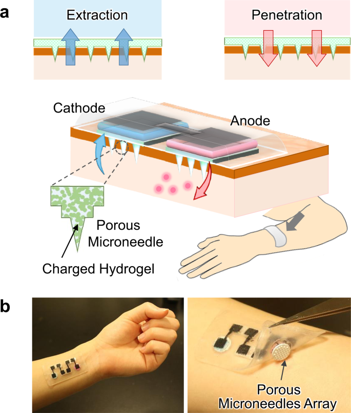
In this study, we succeeded in the generation of a larger transdermal EOF by using PMN, aiming at applications for efficient drug delivery (penetration) and analysis of ISF (extraction), as illustrated in Fig. 1a. The ion-conductive PMN significantly lowered the transdermal resistance by partial breaking of the stratum corneum, and its modification with a hydrogel containing sulfonic groups realized the generation of the transdermal EOF in the anode-to-cathode direction.
The PMN-generated EOF enhanced the transdermal molecular penetration or extraction, similarly to the flow induced by external pressure. We employed modification of the negatively charged hydrogel according to the polarity of the skin tissue. The higher the density of sulfonic groups in the hydrogel, the larger was the flow velocity of the transdermal EOF.
The EOF-assisted delivery of a model drug (fluorescently labeled dextran, 10,000 Da) and the extraction of glucose was demonstrated using a pig skin sample. The driving of the transdermal EOF system with an enzymatic biobattery (fructose/O2 biobattery) was also demonstrated to explore the possible construction of a totally organic EOF patch (Fig. 1b).
More information: Shinya Kusama et al. Transdermal electroosmotic flow generated by a porous microneedle array patch, Nature Communications (2021). DOI: 10.1038/s41467-021-20948-4


