


 Extract (6-MSITC) in Healthy Older Adults")
: An In-Depth Exploration into its Thermogenic Role and Social Significance")


In a major collaborative effort, researchers at the Lewis Katz School of Medicine at Temple University and the University of Nebraska Medical Center (UNMC) have for the first time eliminated replication-competent HIV-1 DNA – the virus responsible for AIDS – from the genomes of living animals.
The study, reported online July 2 in the journal Nature Communications, marks a critical step toward the development of a possible cure for human HIV infection.
“Our study shows that treatment to suppress HIV replication and gene editing therapy, when given sequentially, can eliminate HIV from cells and organs of infected animals,” said Kamel Khalili, Ph.D., Laura H. Carnell Professor and Chair of the Department of Neuroscience, Director of the Center for Neurovirology, and Director of the Comprehensive NeuroAIDS Center at the Lewis Katz School of Medicine at Temple University (LKSOM). Dr. Khalili and Howard Gendelman, MD, Margaret R. Larson Professor of Infectious Diseases and Internal Medicine, Chair of the Department of Pharmacology and Experimental Neuroscience and Director of the Center for Neurodegenerative Diseases at UNMC, were senior investigators on the new study.
“This achievement could not have been possible without an extraordinary team effort that included virologists, immunologists, molecular biologists, pharmacologists, and pharmaceutical experts,” Dr. Gendelman said.
“Only by pooling our resources together were we able to make this groundbreaking discovery.”
Current HIV treatment focuses on the use of antiretroviral therapy (ART).
ART suppresses HIV replication but does not eliminate the virus from the body.
Therefore, ART is not a cure for HIV, and it requires life-long use. If it is stopped, HIV rebounds, renewing replication and fueling the development of AIDS.
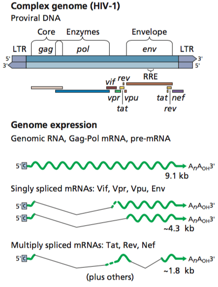
HIV rebound is directly attributed to the ability of the virus to integrate its DNA sequence into the genomes of cells of the immune system, where it lies dormant and beyond the reach of antiretroviral drugs.
In previous work, Dr. Khalili’s team used CRISPR-Cas9 technology to develop a novel gene editing and gene therapy delivery system aimed at removing HIV DNA from genomes harboring the virus.
In rats and mice, they showed that the gene editing system could effectively excise large fragments of HIV DNA from infected cells, significantly impacting viral gene expression. Similar to ART, however, gene editing cannot completely eliminate HIV on its own.
For the new study, Dr. Khalili and colleagues combined their gene editing system with a recently developed therapeutic strategy known as long-acting slow-effective release (LASER) ART. LASER ART was co-developed by Dr. Gendelman and Benson Edagwa, Ph.D., Assistant Professor of Pharmacology at UNMC.
LASER ART targets viral sanctuaries and maintains HIV replication at low levels for extended periods of time, reducing the frequency of ART administration.
The long-lasting medications were made possible by pharmacological changes in the chemical structure of the antiretroviral drugs.
The modified drug was packaged into nanocrystals, which readily distribute to tissues where HIV is likely to be lying dormant.
From there, the nanocrystals, stored within cells for weeks, slowly release the drug.
According to Dr. Khalili, “We wanted to see whether LASER ART could suppress HIV replication long enough for CRISPR-Cas9 to completely rid cells of viral DNA.”
To test their idea, the researchers used mice engineered to produce human T cells susceptible to HIV infection, permitting long-term viral infection and ART-induced latency.
Once infection was established, mice were treated with LASER ART and subsequently with CRISPR-Cas9.
At the end of the treatment period, mice were examined for viral load.
Analyses revealed complete elimination of HIV DNA in about one-third of HIV-infected mice.
“The big message of this work is that it takes both CRISPR-Cas9 and virus suppression through a method such as LASER ART, administered together, to produce a cure for HIV infection,” Dr. Khalili said.
“We now have a clear path to move ahead to trials in non-human primates and possibly clinical trials in human patients within the year.”
Although antiretroviral therapy (ART) has been highly effective at controlling HIV-1 viral loads in the bloodstream of infected individuals, the virus remains latent in infected cells and starts replicating within a couple of weeks upon termination of therapy.
The discovery of CRISPR/Cas9 gene editing technology has provided renewed hope for alternative suppressive therapies and possible elimination of HIV-1 integrated proviral DNA from the genomes of infected individuals.
However, even though this strategy has been somewhat successful in in vitro laboratory cell models, editing of HIV-1 DNA in cells and tissues that support viral reservoirs in vivo has been challenging.
Accordingly, recent strategies have employed targeted vectors to deliver CRISPR/Cas9 complexes directly to infected subjects.
In a recent study at Temple University, the investigators sought to excise HIV-1 DNA from patient immune cell engrafts in mice by treating the mice with a CRISPR/Cas9 complex delivered in a lentiviral vector cocktail.
The mice were immunodeficient animals that were “humanized” by injection with blood cells from healthy or HIV-1-positive human subjects. The cocktail (“therapeutic cocktail”) consisted of three separate lentiviruses encoding either
a) a CRISPR guide RNA targeting the 5’ HIV-1 long terminal repeat (LTR) motif;
b) a CRISPR guide RNA targeting the 3’ HIV-1 LTR motif; or
c) a Cas9 effector protein.
This cocktail would facilitate cleavage of integrated HIV-1 DNA on both 5’ and 3’ LTRs, resulting in the excision of a portion of each LTR and the entire HIV genome positioned between the two LTRs, corresponding to the gag-pol-env and accessory genes.
The experimental design, which aimed to assess the combined ability of the individual components in this therapeutic cocktail to edit the HIV-1 genome in vitro or in vivo, was as follows.
1. In vitro HIV-1 infection of healthy human blood cells, followed by in vitro treatment with therapeutic cocktail.
Blood cells isolated from healthy human subjects were infected with HIV-1 and treated with the therapeutic cocktail one week post-infection.
The efficiency of excision of HIV-1 DNA was determined by sequencing the 5’ and 3’ LTR regions. There was a significant reduction (up to 65%) of HIV-1 DNA in treated cells versus non-treated control cells.
2. Engraftment of human in vitro infected HIV-1 positive cells into mice, followed by treatment with therapeutic cocktail in vivo.
Human HIV-1-positive blood cells infected in vitro were injected into immunodeficient mice to generate humanized mice.
One week later, the mice were treated with the therapeutic cocktail.
After another week, the mice were euthanized and the efficiency of excision of HIV-1 DNA was determined. In agreement with the results obtained in step 1, there was a drastic decline in HIV-1 DNA in engrafted treated animals versus engrafted non-treated control animals.
3. Ex vivo treatment of human blood cells derived from HIV-1-positive donors, with therapeutic cocktail.
Blood cells were isolated from three separate HIV-1-positive patients (patients 1-3), each of whom were on ART therapy and showed low or non-detectable virus levels.
Their cells were treated with the therapeutic cocktail, and the efficiency of excision of HIV-1 DNA was determined.
here was a significant decrease in the level of HIV-1 DNA (up to 68%) in treated cells versus non-treated control cells.
4. Engraftment of human blood cells from HIV-1-positive donors into mice, followed by treatment with therapeutic cocktail in vivo.
Blood cells from patients 1-3 were injected into immunodeficient mice to generate humanized mice. One week later, the mice were treated with the therapeutic cocktail.
Two weeks later, the mice were euthanized and the efficiency of excision of HIV-1 DNA in the blood, spleens, and various other organs was determined.
The efficiency of excision observed in the blood and spleens of mice engrafted with cells from patients 1 or 2 was as high as 96% compared to that observed in the blood and spleens of non-treated control mice.
In contrast, the efficiency of excision was as low as 25% in mice engrafted with cells from patient 3.
This difference in efficiency was attributed to variabilities observed in viral LTR sequences and guide RNA target sequences between different patient samples.
As a further assessment of the efficacy of this therapeutic strategy, the authors isolated cells from the blood and spleen of therapeutically treated mice that were engrafted with cells from patient 1 and co-cultured these cells with a cell line that is highly permissive for HIV-1 infection.
They observed significantly reduced recovery of HIV-1 from cells taken from engrafted treated animals versus engrafted non-treated animals.
Although these results are promising, the system is still artificial, making it difficult to predict if it would work in humans.
Furthermore, in vivo editing of the HIV-1 genome by CRISPR/Cas9 may not completely remove replication-competent virus from organs serving as reservoirs for viral latency, such as the spleen. A
s explained by John Coffin and Stephen Hughes on TWiV 510, the viral reservoir exists in a pool of infected cells that contain HIV integrated provirus.
These cells are capable of clonally expanding, and surprisingly, not all offspring of a clone exhibit identical levels of viral expression.
Developing effective strategies to identify and eliminate such pools of cells is a prevailing challenge in this field. Likely, complete elimination of HIV-1 by CRISPR may only be accomplished in combination with other therapies, such as ART, and may require personalized CRISPR/Cas9 complexes that are perfectly matched to a particular viral genome.
More information: Sequential LASER ART and CRISPR Treatments Eliminate HIV-1 in a Subset of Infected Humanized Mice, Nature Communications (2019). DOI: 10.1038/s41467-019-10366-y , https://nature.com/articles/s41467-019-10366-y
Journal information: Nature Communications
Provided by Temple University


