


 Extract (6-MSITC) in Healthy Older Adults")
: An In-Depth Exploration into its Thermogenic Role and Social Significance")


Researchers from Carnegie Mellon University (CMU) and Nanyang Technological University, Singapore (NTU Singapore) have developed an organ-on-an-electronic-chip platform, which uses bioelectrical sensors to measure the electrophysiology of the heart cells in three dimensions.
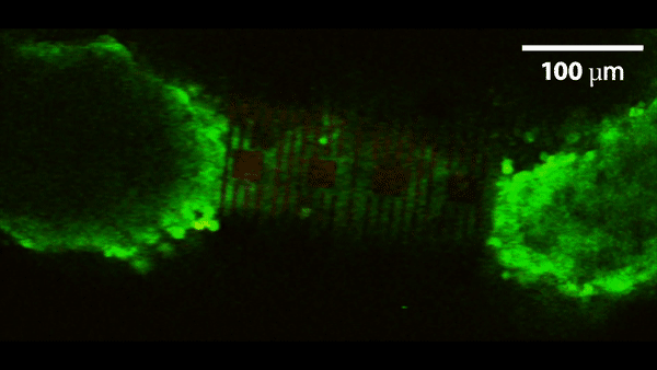
These 3-D, self-rolling biosensor arrays coil up over heart cell spheroid tissues to form an “organ-on-e-chip,” thus enabling the researchers to study how cells communicate with each other in multicellular systems such as the heart.
The organ-on-e-chip approach will help develop and assess the efficacy of drugs for disease treatment—perhaps even enabling researchers to screen for drugs and toxins directly on a human-like tissue, rather than testing on animal tissue.
The platform will also be used to shed light on the connection between the heart’s electrical signals and disease, such as arrhythmias.
The research, published in Science Advances, allows the researchers to investigate processes in cultured cells that currently are not accessible, such as tissue development and cell maturation.
“For decades, electrophysiology was done using cells and cultures on two-dimensional surfaces, such as culture dishes,” says Associate Professor of Biomedical Engineering (BME) and Materials Science & Engineering (MSE) Tzahi Cohen-Karni.
“We are trying to circumvent the challenge of reading the heart’s electrical patterns in 3-D by developing a way to shrink-wrap sensors around heart cells and extracting electrophysiological information from this tissue.”
The “organ-on-e-chip” platform starts out as a small, flat rectangle, not unlike a microscale slap bracelet.
A slap bracelet starts out as a rigid, ruler-like structure, but when you release the tension it quickly coils up to band around the wrist.
The organ-on-e-chip starts out similarly.
The researchers pin an array of sensors made of either metallic electrodes or graphene sensors to the chip’s surface, then etch off a bottom layer of germanium, which is known as the “sacrificial layer.”
Once this sacrificial layer is removed, the biosensor array is released from its hold and rolls up from the surface in a barrel shaped structure.
The researchers tested the platform on cardiac spheroids, or elongated organoids made of heart cells.
These 3-D heart spheroids are about the width of 2-3 human hairs. Coiling the platform over the spheroid allows the researchers to collect electrical signal readings with high precision.
“Essentially, we have created 3-D self-rolling biosensor arrays for exploring the electrophysiology of induced pluripotent stem cell derived cardiomyocytes,” says lead author of the study and BME Ph.D. student Anna Kalmykov.
“This platform could be used to do research into cardiac tissue regeneration and maturation that potentially can be used to treat damaged tissue after a heart attack, for example, or developing new drugs to treat disease.”
Through collaboration with the labs of BME/MSE Professor Adam Feinberg and former CMU faculty Jimmy Hsia, now Dean of the Graduate College of NTU Singapore, the researchers were able to design a proof of concept and test them on 3-D micro-mold formed cardiomyocyte spheroids.

“Mechanics analysis of the roll-up process enables us to precisely control the shape of the sensors to ensure conforming contact between the sensors and the cardiac tissue,” says NTU Professor Jimmy Hsia.
“The technique also automatically adjusts the level of the delicate ‘touch’ between the sensors and the tissue such that high quality electric signals are measured without changing in the physiological conditions of the tissue due to external pressure.”
“The whole idea is to take methods that are traditionally done in planar geometry and do them in three dimensions,” says Cohen-Karni.
“Our organs are 3-D in nature. For many years, electrophysiology was done using just cells cultured on a 2-D tissue culture dish.
But now, these amazing electrophysiology techniques can be applied to 3-D structures.”
More information: A. Kalmykov el al., “Organ-on-e-chip: Three-dimensional self-rolled biosensor array for electrical interrogations of human electrogenic spheroids,” Science Advances (2019). advances.sciencemag.org/content/5/8/eaax0729
Journal information: Science Advances
Provided by Carnegie Mellon University
What are tissue chips?
Tissue chips, or microphysiological systems (MPS), are devices designed to position cells in a three-dimensional structure that mimic the function of organs of the body, and react in a physiological manner to exposure to drugs, hormones, cell signaling molecules and biomechanical stressors. Platforms vary in design, with some systems allowing cells to self-organize into organoid-type structures, and others providing scaffolding for cells to proliferate and grow in a structurally-defined way.
Some have highly prescriptive designs, where specific cell types are placed in well-defined positions or compartments to recapitulate functional units of organs, such as the kidney proximal tubule or liver sinusoid.
The wide range of designs means a wide range of platform sizes too, ranging from cell compartments of a few hundred micrometers resting on microscope slide-sized platforms, to multi-organ systems with a footprint of a few centimeters.
What all systems have in common is the recapitulation of miniaturized functional units of human organ systems, many thousands or millions of times smaller than the actual organ, and the employment of microfluidic technology to allow for fluid flow through the system to deliver nutrients and remove cellular waste, either by pump or gravity.
Also common to all MPS platforms is the three-dimensional (3D) cellular arrangement of multiple cell types, which allows functional tissue-tissue interfaces and complex cellular communication to be recapitulated in vitro.
These last two design features – fluid flow and 3D spatial cellular arrangements – subject tissues to shear and stretch forces that mimic in vivo conditions and are difficult to model in two dimensions.
Additionally, chips are often designed from clear plastics or materials that enable cells to be visualised through the device via microscopy, allowing for real-time imaging and monitoring of cell function and health over a longer period of time.
This kind of longitudinal monitoring of cell function allows the time course of response and recovery to be modeled, and the effects of cyclical hormone patterns on drug response over time investigated. Additionally, the flow of fluid through the systems allows for collection of platform effluent for further enzymatic or biochemical assays. The wide diversity of platform designs (see Figure 1) allows a broad range of biological questions to be addressed in novel and innovative ways.

A broad array of tissue chip platforms have been developed. These include (clockwise from top right) a blood-brain barrier (Wikswo lab at Vanderbilt University), cardiac muscle (Parker lab at Harvard), kidney proximal tubule (www.nortis.com), female reproductive tract (DRAPER laboratories), vascularized tumor (George lab at Washington University), skin epidermis (Christiano lab at Columbia), vasculature (George lab at Washington University), liver (Taylor lab at University of Pittsburgh), and lung (www.emulatebio.com). Center image from www.ncats.nih.gov/tissuechip. All images reproduced with permission from the developers.
Broadly speaking, the ability to recreate functional human (and animal) organs in vitro could transform the drug discovery process, and open new avenues for the study of physiology and disease pathology in both humans and animals. This review will discuss the current state of the technology, drug development and disease modeling applications, and challenges faced by the field moving into the future.


