


 Extract (6-MSITC) in Healthy Older Adults")
: An In-Depth Exploration into its Thermogenic Role and Social Significance")


Emphysema is a progressive, debilitating lung disease in which the lung’s breathing sacs, or alveoli, enlarge, get thinner, and eventually are destroyed as the cells die off.
It can be fatal, and there is currently no cure. New research at Boston Children’s Hospital, using lung cells and mouse models of emphysema, offers hope in the form of a small, engineered peptide molecule called PR1P.
Findings were published July 14 in the American Journal of Respiratory Cell and Molecular Biology.
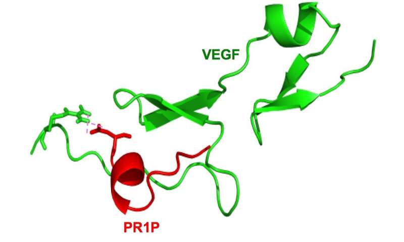
The peptide was developed by Avner Adini, Ph.D., Robert D’Amato, MD, Ph.D., Benjamin Matthews, MD, and other colleagues in Boston Children’s Vascular Biology Program, based on previous research.
That research had found that vascular endothelial growth factor (VEGF), known to be key for blood vessel development, also helps to keep lung tissue healthy, and that VEGF signaling goes awry in emphysema, hastening cell death.
“VEGF is found at high levels in the normal lung – more than 500 times the levels seen in blood plasma,” says Adini.
“VEGF levels tend to become dramatically disrupted during lung disease, but return to normal during recovery. It is not clear whether VEGF dysregulation is the root cause of lung disease, or the result of the lung pathology. In emphysema, VEGF activity is low.”
Maintaining VEGF levels in the lung
The scientists’ first thought was to try to give VEGF directly as a therapy, but VEGF has a very short half-life and can potentially produce serious side effects, including low blood pressure, in animal models.
“So we asked, could there be an alternative way to upregulate VEGF signaling to make it more potent, rather than directly boost VEGF concentrations in the lung?” says Adini.
Serendipitously, through previous research on melanoma, Adini discovered a protein called prominin-1 bound directly to VEGF.
Using molecular biology and genetic engineering approaches, he and his colleagues fashioned PR1P, a small peptide comprised of the same 12 amino acids that prominin-1 uses to bind to VEGF.
Protecting the lungs in emphysema models
When the team tested PR1P in lung cells and mouse models of emphysema, they found that it protected VEGF from being degraded. In fact, mice that inhaled PR1P had significantly increased VEGF levels in their lungs as early as 30 minutes after treatment. VEGF levels remained twice as high as in control animals 24 hours later.
Importantly, the researchers further found that PR1P reduced apoptosis (programmed cell death, a key feature in emphysema) in both cell cultures and in live mice.
Finally, they showed that PR1P reduced lung damage in mice with toxin-induced emphysema that were followed for 4 and 21 days. Although the study was not designed to assess toxicity, they did not observe any adverse effects.
In addition to emphysema, PR1P may have therapeutic potential for acute respiratory distress syndrome (ARDS), a common and devastating disease which also has no cure and is also characterized by abnormalities in VEGF signaling.
Adini and Matthews have funding from the Boston Biomedical Innovation Center (B-BIC) to support their ongoing work to develop PR1P as a therapeutic for both emphysema and ARDS.
“We are now also seeing dramatic lung-preserving and anti-inflammatory effects of inhaled PR1P in mice exposed to multiple diverse toxins that induce lung injury in ARDS models,” says Adini.
Cancer is one of the leading causes of death worldwide. The incidence and mortality rates of cancer have tremendously increased, constituting an enormous burden on public health, cancer patients and their families, and society as a whole (Torre et al., 2016; Gonzaga et al., 2018).
Therefore, various strategies in cancer therapy to improve therapeutic efficacy have been developed in preclinical and clinical settings recently. These treatment strategies mainly include novel therapeutic targets, such as antiangiogenesis, apoptosis-related signaling pathways, oncogene inactivation, and tumor suppressors and immunotherapy (Cross and Burmester, 2006).
Among these strategies, induction of apoptosis and repression of angiogenesis are considered two promising approaches in cancer therapy (Ding and Fisher, 2002; Carmeliet and Jain, 2011; Potente et al., 2011). For example, sorafenib, a small molecule inhibitor of several kinases including Raf, vascular endothelial growth-factor receptors (VEGFR) and platelet-derived growth factor receptor (PDGFR) involved in the proliferation and angiogenesis of cancer cells, showed significantly therapeutic efficacy in the treatment of patients with various cancers, including liver, lung, and colorectal cancers clinically (Rini, 2006; Blumenschein, 2008; Mellema et al., 2013; Ogasawara et al., 2017).
Angiogenesis plays a crucial role in tumor growth by ensuring the supply of sufficient oxygen and fundamental nutrients to cancer cells and tissues (Tabata, 2003; Zhu et al., 2010).
Angiogenesis is regulated by various pro-angiogenic factors such as VEGF, PDGF, basic fibroblast growth factor (bFGF), transforming growth factor-beta (TGF-β), angiogenin, and interleukin-8 (IL-8) ect. These factors promote the stimulation, proliferation, and invasion of endothelial cells, resulting in the excessive growth of new capillaries into the tumor (Verheul et al., 2004; Kerbel, 2008).
VEGF is one of the most important regulators of angiogenesis (Shibuya, 2001). VEGF is secreted from cancer and stromal cells in the tumor microenvironment for promoting the growth and migration of vascular endothelial cells and the permeability of blood vessels (Ferrara, 2004).
Moreover, several key intracellular signaling molecules including phosphoinositide 3-kinase (PI3K), protein kinase B (AKT), and mammalian target of rapamycin (mTOR) are activated when VEGF binds to VEGFR2 on endothelial cells, which are responsible for the proliferation, migration, invasion, and survival of endothelial cells (Stefanini et al., 2009; Kim et al., 2013; Jiao et al., 2016; Kim, 2017). Therefore, anti-VEGF therapy has clearly demonstrated antitumor efficacy via inhibition of angiogenesis against various malignancies clinically (Grothey and Galanis, 2009).
Aberrant apoptosis is a major reason for cancer development, survival, and progression (Lowe and Lin, 2000; Tayyaba et al., 2016). The ability to evade apoptosis is an important feature of cancer cells. Bcl-2 and Bax belong to the Bcl-2 family, which are the most important apoptosis regulatory molecules (Liu et al., 2011; Yao et al., 2017). Bcl-2 and Bax play important roles in the mitochondrial apoptotic pathway, with both factors having opposing functions (Liang et al., 2016).
The ratio of Bcl-2 and Bax affects the relative sensitivity or resistance of cancer cells to apoptotic stimuli and therapeutic drugs (Liu et al., 2011). Caspase-3, a downstream effector molecule, is a proteolytic enzyme that executes apoptotic cell death. Therefore, apoptosis is a key target for cancer therapy.
Sanguisorba officinalis L. is a traditional Chinese herb that is widely used for immunomodulation and treatment of blood toxicity, hepatitis B, and cancer (Kim et al., 2001; Cai et al., 2012; Wang et al., 2012; Yang et al., 2015; Liu et al., 2016). Tannin, one of the main components of Sanguisorba officinalis L., exhibits antibiotic, antiviral, and hematopoietic effects (Sharma et al., 2011; Adini et al., 2017).
Recent pharmacological studies have shown that tannin could inhibit the growth of breast cancer cells and angiogenesis of human umbilical vein endothelial cells (HUVECs) (Wang et al., 2012).
Moreover, previous study revealed that ellagic acid suppressed angiogenesis in HUVECs and exhibited antitumor activity against sarcoma S180 and liver cancer H22 (Ya et al., 2015). However, the study of the effects of 3,3′,4′-trimethylellagic acid (TMEA, an ellagic acid) on the anticancer activity and angiogenesis is limited.
To determine the antitumor effects of TMEA, the cell proliferation was determined by MTS and the mRNA and protein expressions of Bcl-2, Bax, and caspase-3 in liver cancer HepG2, lung cancer A549, and colon cancer SW620 cells by qRT-PCR and Western blotting analysis, respectively.
Furthermore, the antitumor activity of TMEA was evaluated in SW620 tumor xenograft bearing in nude mice in vivo and the expressions of CD31, Bcl-2, Bax, and caspase-3 were investigated in SW620 tumor tissues by immunohistochemical analysis. In addition, the effects of TMEA on molecular docking with VEGFR2, VEGF expression, and VEGF-induced angiogenesis were investigated by wound healing and tube formation assay in HUVECs.
More information: Avner Adini et al, PR1P Stabilizes VEGF and Upregulates its Signaling to Reduce Elastase Induced Murine Emphysema, American Journal of Respiratory Cell and Molecular Biology (2020). DOI: 10.1165/rcmb.2019-0434OC


