


 Extract (6-MSITC) in Healthy Older Adults")
: An In-Depth Exploration into its Thermogenic Role and Social Significance")


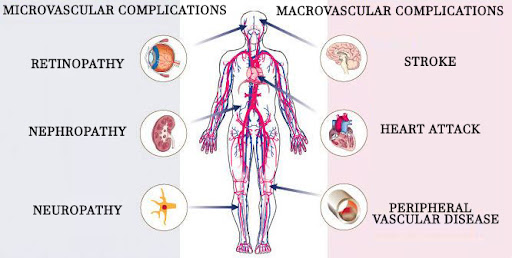
A new study by researchers from the University of Texas-Arlington shows that young and otherwise healthy adults would suffer persistent peripheral macrovascular and microvascular complications following ‘recovery’ from COVID-19.
The study was the first to investigate the persistent effects of COVID-19 on vascular function in otherwise healthy young adults.
The study findings demonstrated that peripheral macrovascular and microvascular vasodilation was significantly blunted in young adults still symptomatic from COVID-19 beyond the acute phase (>4 wk from diagnosis), whereas those who become asymptomatic have similar vascular function compared with controls who never had COVID-19.
In contrast, cerebral vascular function and central arterial stiffness were unaffected irrespective of COVID-19 symptomology.
Numerous past studies suggest that COVID-19 causes vascular dysfunction during the acute phase of the illness in otherwise healthy young adults. T
o date no studies have investigated the longer-term effects of COVID-19 on vascular function.
The study team hypothesized that young, otherwise healthy adults who are past the acute phase of COVID-19 would exhibit blunted peripheral (brachial artery flow-mediated dilation (FMD) and reactive hyperemia) and cerebral vasodilator function (cerebral vasomotor reactivity to hypercapnia; CVMR) and increased central arterial stiffness.
A total of sixteen young adults who were at least 4 wk past a COVID-19 diagnosis and 12 controls who never had COVID-19 were studied. Eight subjects with COVID-19 were symptomatic (SYM) and eight were asymptomatic (ASYM) at the time of testing.
The study findings found that FMD and reactive hyperemia were not different between COVID and control groups. However, FMD was lower in SYM (3.8 ± 0.6%) compared with ASYM (6.8 ± 0.9%; P = 0.007) and control (6.8 ± 0.6%; P = 0.003) with no difference between ASYM and control.
Similarly, peak blood velocity following cuff release was lower in SYM (47 ± 8 cm/s) compared with ASYM (64 ± 19 cm/s; P = 0.025) and control (61 ± 14 cm/s; P = 0.036). CVMR and arterial stiffness were not different between any groups.
In summary, peripheral macrovascular and microvascular function, but not cerebral vascular function or central arterial stiffness were blunted in young adults symptomatic beyond the acute phase of COVID-19. In contrast, those who were asymptomatic had similar vascular function compared with controls who never had COVID-19.
The study findings were published in the peer reviewed journal: Heart and Circulatory Physiology (American Journal Of Physiology )
https://journals.physiology.org/doi/full/10.1152/ajpheart.00368.2021
Lead author Dr Damsara Nandadeva from the Department of Kinesiology, University of Texas at Arlington told Thailand Medical News, “It is intriguing that those with persistent [COVID-19] symptoms exhibited peripheral vascular dysfunction, whereas those who were asymptomatic at the time of testing had similar macrovascular and microvascular vasodilation to controls.”
It should be noted that adults 29 years of age and younger makeup only 16% of the U.S. population but account for over 7 million, or 22%, of U.S. COVID-19 infections.
Although more research is needed to fully understand the extent of the problem, one study found that as many as 1 in 4 adults ages 18 to 39 reported symptoms weeks or months after the initial acute phase of COVID-19 infection.
Dr Nandadeva explained, “Long COVID,” also called post-acute sequelae of SARS-COV-2 infection (PASC), is an umbrella term for the condition the U.S. Centers for Disease Control and Prevention refers to as “a lack of return to a usual state of health following acute COVID-19 illness.”
It has been found that individuals with long COVID report a variety of symptoms, including cognitive impairment, headache, and lightheadedness, which can be indicative of impairments to blood vessels supporting the brain.
The study team from the University of Texas at Arlington investigated brai
n and peripheral blood vessel function of 16 young adults who had tested positive for COVID-19 more than four weeks prior. Eight of the study subjects currently had COVID-19 symptoms and eight did not. Researchers then compared them to 12 peers who tested negative.
Interestingly, symptomatic participants showed “significantly blunted” dilation of both large and small blood vessels in their limbs. However, despite previous studies showing stiffening of central arteries in young adults with acute COVID-19, long COVID participants in this study showed no such stiffening, nor did they show impaired function in the blood vessels that support the brain.
Also it was observed that asymptomatic COVID-19–positive participants showed function similar to controls, offering hope that once symptoms resolve so may vascular impairments.
However during these vulnerable phases, affected individuals should constantly be monitored and should go for frequent checkups to ensure no serious or fatal events arises.
SARS-CoV-2 infection and vascular dysfunction
In health, the vascular endothelium maintains homeostasis through regulation of immune competence, inflammatory equilibrium, tight junctional barriers, hemodynamic stability as well as optimally balanced thrombotic and fibrinolytic pathways. In the novel coronavirus disease of 2019 (COVID-19) caused by the severe acute respiratory syndrome coronavirus 2 (SARS-CoV-2), dysregulation of many of these pathways has emerged as a mediator of severe disease.
The constellation of clinical and biomarker derangements seen in COVID-19 can be classified into disruption of the immune, renin-angiotensin-aldosterone (RAA), and thrombotic balance, all of which converge on the vascular endothelium as a common pathway.
Accumulating evidence from basic science, imaging and clinical observations, has clarified the picture of COVID-19 as a vascular disease. Understanding the disease in this context may provide novel avenues of understanding COVID-19 and lead to critically needed improvements in therapeutic strategies.
SARS-CoV-2 uses the angiotensin converting enzyme 2 (ACE2) to facilitate entry into target cells and initiate infection. This viral entry into the cell is further mediated by transmembrane serine protease 2 (TMPRSS2) and cathepsin L which cleave the S protein on the viral particle to permit engagement with ACE2 [1]. Endothelial cells (ECs) in general and cardiac pericytes in particular express abundant ACE2, making them a direct target of SARS-CoV-2 infection (Fig. 1 ) [2].
Examination of the pulmonary vascular bed shows severe derangements in COVID-19, compared to control and influenza patients, particularly with widespread thrombosis and microangiopathy, endothelial activation and extensive angiogenesis [3]. These studies and pervasive findings establish the role of viral injury to the vascular system with resulting vascular dysfunction in COVID-19 patients [4].

SARS-CoV-2 Induced Endothelial Injury
Legend: A schematic of SARS-CoV-2 infection and proposed resulting endothelial injury, involving immune activation, pro-thrombotic milieu, and RAAS dysregulation. These insults interact with each other to cause end-organ dysfunction that is manifest in many COVID-19 patients.
TMPRSS2 = Transmembrane protease serine 2; ADAM17 = A disintegrin and metalloproteinase 17; TNF = Tumor necrosis factor; TNFr = Tumor necrosis factor receptor; TLR = toll-like receptor; DAMPs = Damage-associated molecular patterns; PAMPs = Pathogen-associated molecular patterns; PAI-1 = plasminogen activator inhibitor-1; vWF = von Willebrand factor; eNOS = endothelial nitric oxide; tPA = tissue plasminogen activator; AT1R = angiotensin 1 receptor; ARDS = acute respiratory distress syndrome
Created with BioRender.com.
COVID-19, immune dysregulation and endothelial injury
The vascular endothelium has an intricate role in immune regulation and inflammation, an axis that SARS-CoV-2 infection disturbs. Reports from hospitalized COVID-19 patients reveal an activation of the immune system that displays findings consistent with cytokine storm, macrophage activating syndrome and subsequent immune exhaustion, most severe in the sickest patients [5,6].
This hyper-inflammatory state has deleterious effects on the vascular system with resulting EC dysfunction. In the presence of circulating inflammatory mediators such as interleukin (IL)-1, IL-6, damage-associated molecular patterns and pathogen-associated molecular patterns, ECs undergo transition to an activated state that participates in host defenses [7].
Activated ECs promote localized inflammation by inducing proinflammatory gene expression, attracting immune cells, promoting recruitment of inflammatory cells to injured or infected tissues, vascular leak by increasing endothelial permeability, and altering the thrombotic potential of the local intimal surface.
Activation of neutrophils leads to formation of neutrophil extracellular traps (NETs), a process sometimes referred to as NETosis, which may contribute to responses to pathogens and thrombosis [8]. Additionally, EC injury is compounded by toll-like receptor (TLR) activation by viral RNA recognition, with resulting increased reactive oxidative species (ROS) production [9].
In a regulated and self-limited immune response, these mechanisms help to quell the local insult, with subsequent healing and return to a resting EC state. In contrast, states of a heightened innate immune response and a prothrombotic state elicited by innate immune mediators such as that seen in COVID-19 lead to an escalating cascade of these pathways that promote profound micro- and macrovascular EC dysfunction and damage, with impairment of other important functions of the endothelium (Fig. 1).
Recent reports of a hyperinflammatory syndrome similar to Kawasaki disease shock syndrome in children, with elevated biomarkers of cardiac injury, development of arrythmia and progression to giant coronary aneurysms in some cases further sheds light on one manifestation of a macrovascular-focused immune phenomenon in COVID-19, an area that warrants further study [10].
The EC dysregulation imposed by COVID-19 extends beyond the circulatory system. Pulmonary ECs have an important role in immune surveillance, maintaining alveolar integrity and ensuring appropriate oxygen exchange. In COVID-19, direct viral infection and the systemic inflammatory response likely lead to severe dysfunction of these important ECs, compounding the resulting picture of severe hypoxia and acute respiratory distress syndrome frequently reported in hospitalized patients.
Endothelial injury and thrombosis in COVID-19
The endothelium, along with its key immunoregulatory functions, also plays an essential role in maintaining a dynamic interplay between the pro-coagulant and fibrinolytic factors in the vascular system. In the quiescent state, the endothelium forms a barrier between the prothrombotic subendothelial layer and pro-coagulant factors in the blood.
In times of stress, activated ECs express more plasminogen activator inhibitor-1 (a key inhibitor of endogenous fibrinolysis), tissue factor (a potent procoagulant) and release von Willebrand factor (a protein that forms multimers that promote thrombus growth).
Activated ECs lower activity of thrombomodulin and tissue plasminogen activator, favoring thrombus accumulation [11]. Furthermore, during stress, immune effectors potentiate the thrombotic response, including NET formation, and thus can also propagate thrombosis [8].
These changes, along with other complex regulatory pathways lead to endothelial dysfunction, activation of the coagulation cascade and suppression of fibrinolytic mechanisms, with a resulting prothrombotic milieu (Fig. 1) [11]. Such disturbances in the fine balance of endothelial health and the coagulation and fibrinolytic systems lead to both macrovascular and diffuse microvascular thrombotic disease in the patient [12].
Clinical studies show that patients with COVID-19 have increased fibrinogen, fibrin degradation products, D-dimer and von Willebrand factor, and these elevations appear to correlate with severity of disease and thrombotic risk [13,14]. Early reports showed a substantial burden of myocardial injury in patients who were critically ill or died from COVID-19 [15].
Multiple mechanisms may cause cardiac injury in COVID-19 [16]. While infection and hemodynamic stresses of acute critical illness can trigger plaque rupture and resulting myocardial infarction, recent reports indicate that some COVID-19 patients show biomarker and electrocardiographic findings of myocardial infarction without evidence of acute plaque rupture on angiography [17].
Furthermore, a recent case series reported a 78% prevalence of cardiovascular involvement and myocardial inflammation without apparent left ventricular impairment on cardiac magnetic resonance imaging in recovered COVID-19 patients without cardiac symptoms post-discharge from the hospital [18]. This pattern of cardiac injury could result from endothelial dysfunction and coronary microvascular thrombosis in these patients, rather than coronary macrovascular thrombosis.
Evidence of endothelial injury also comes from autopsy reports that provide further support of this pan-vascular involvement in COVID-19, with series showing compelling evidence of a high incidence of both deep venous thrombosis as well as in situ pulmonary arterial thrombosis [3,19].
Prominent findings in these autopsies included microvascular thrombi, particularly in the pulmonary circulation, pointing to a disrupted endothelium with a resultingly pro-coagulable state in COVID-19. Multiple analyses and systematic reviews have shown a substantial burden of venous thromboembolism (VTE) in COVID-19 patients, with up to 25% incidence of VTE and 20% incidence of pulmonary embolism in hospitalized patients, particularly those with more severe illness requiring critical care and amongst those not on prophylactic or therapeutic anticoagulation [14,[20], [21], [22]].
Further support for macrovascular manifestations of endothelial dysfunction with an activation of pro-thrombotic mechanisms comes from reports highlighting the higher incidence of large-vessel stroke in COVID-19 patients as well as well as a retrospective case-control analysis showing COVID-19 to be an independent risk factor for stroke [23,24].
Recent obstetric data also add evidence to SARS-CoV-2 acting through endothelial and vascular mechanisms. In a case series of 42 pregnancies admitted with SARS-CoV-2 infection, 14% of all women and 75% of women with severe COVID-19 symptoms manifested a constellation of symptoms similar to pre-eclampsia and hemolysis, elevated liver enzymes, low platelet count (HELLP) syndrome, with spontaneous resolution after COVID-19 recovery [25]. Pre-eclampsia has considerable vascular and endothelial dysfunction in its central pathobiology, and the development of this disorder at a considerable rate in women with COVID-19 suggests endothelial involvement as a sequela of viral infection.
COVID-19 and the renin-angiotensin-aldosterone system
The renin-angiotensin-aldosterone (RAA) system that crucially regulates vascular health and function also participates in COVID-19. As ACE2 contributes critically to the biology of SARS-CoV-2 infection, much attention has focused on the interplay between COVID-19 and the RAA system.
Angiotensin II is the main vascular effector of the RAA system and exerts its deleterious effects on the cardiovascular system via angiotensin-II type 1 (AT1) receptors by activating vasoconstrictor, inflammatory and fibrotic pathways. Prior studies show that angiotensin II perturbs endothelial functions in multiple ways, including by monocyte recruitment, formation of ROS, activation of pro-inflammatory pathways including through nuclear factor kappa-light-chain-enhancer of activated B cells (NF-κB) regulation, as well as promoting plasminogen activator inhibitor-1 (PAI-1) production in ECs [26,27].
In health, ACE2 plays a counter-regulatory role in the RAA system by cleaving angiotensin II to angiotensin 1-7. The enzyme also acts on angiotensin I to convert it to angiotensin 1-9. Both of these enzymatic conversions yield peptides that have potent anti-inflammatory, anti-oxidant and anti-fibrotic properties.
Early data indicate that SARS-CoV-2 limits ACE2 expression by promoting cleavage ACE2 by the specialized proteinase A disintegrin and metalloproteinase 17 (ADAM17) and shedding from the cell surface, leading to a reduction in ACE2’s protective role on ECs and other organs [28].
One study in COVID-19 patients showed a markedly elevated level of serum angiotensin II, further implicating an activated RAA system in the manifestations of SARS-CoV-2 infection [29]. One mechanism by which SARS-CoV-2 infection may affect vascular dysfunction is through oxidative stress.
Nox2, regulated by angiotensin-II contributes to oxidative stress in the endothelium via production of reactive oxidant species, and increases in severe COVID-19, adding to the potential deleterious effects of RAA dysregulation in SARS-CoV-2 infection [30]. Overall, SARS-CoV-2 infection results in RAA system activation with its resulting profile of injurious endothelial functions (Fig. 1). This dysregulation potentiates the innate immune stimulation, oxidative stress and pro-thrombotic states previously described and fuels a vicious cycle in the vascular disease of COVID-19 [26].
reference link : https://www.ncbi.nlm.nih.gov/pmc/articles/PMC7556303/


[…] Majority Of Young Healthy Adults Will Exhibit Peripheral Macrovascular And Microvascular… […]