


 Extract (6-MSITC) in Healthy Older Adults")
: An In-Depth Exploration into its Thermogenic Role and Social Significance")


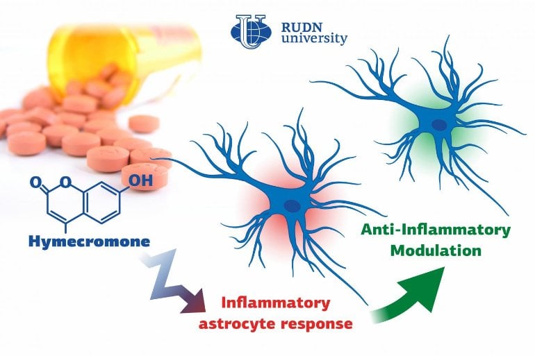
Biologists from RUDN University confirmed that a well-known spasmolytic drug called hymecromone can suppress the inflammatory response in astrocytes, important glial cells of the central nervous system.
All pathological processes in the nervous system, such as neurodegenerative diseases, injuries, or intoxications, are associated with inflammations. It is due to the reaction of many auxiliary cells, including astrocytes that support the neural activity in the brain.
A team of biologists from RUDN University together with their colleagues from Lomonosov Moscow State University confirmed that the intensity of inflammation can be brought down by limiting the number of anti-inflammatory molecules produced by astrocytes.
To do so, they suggested using hymecromone, a drug that is currently used to treat spasms.
“Astrocytes can synthesize hyaluronic acid, one of the main components of the brain’s extracellular matrix. Our studies demonstrated that when hyaluronic acid is added to the astrocytes, it affects the production of the main anti-inflammatory molecules.
In this work, we tested hymecromone that suppresses the synthesis of hyaluronic acid and measured its effect on the inflammatory response of astrocytes,” said Dmitry Chistyakov, PhD, a senior researcher at the Center for Collective Use (Research and Education Center) at RUDN University, and a researcher at Lomonosov Moscow State University.
The team studied the effect of hymecromone on astrocytes in a rat brain cell culture. Cell culture samples were treated with a liposaccharide that activated the receptors in charge of the inflammation, and then with hymecromone. In order to assess the efficiency of the drug, the team extracted RNA from astrocytes and measured the activity of the genes that regulate the production of the anti-inflammatory molecules.
The levels of hyaluronic acid and cytokines were measured in cell supernatants. The changes in the synthesis of signal lipids (oxylipins) were evaluated using the methods of high-efficiency liquid chromatography and tandem mass spectrometry.
Hymecromone turned out to suppress the production of anti-inflammatory molecules: tumor necrosis factor α and interleukins IL-6 and IL-1β, as well as oxylipins. At the same time, it increased the development of IL-10 that helps stop the inflammation. Having measured the levels of hyaluronic acid, the team confirmed that astrocytes actively produced this substance in response to liposaccharides.
However, when a cell culture at the initial stages of the immune response was treated with hymecromone, the levels of hyaluronic acid dropped down. At the same time, hymecromone failed to have the same effect on the cells that did not inflame after contact with the liposaccharide. In these cultures, astrocytes continued to produce hyaluronic acid in regular quantities.
According to the biologists, it is still early to say for sure that the anti-inflammatory properties of hymecromone are rooted in its ability to reduce the production of hyaluronic acid. Still, the team is positive that these processes are connected.
“We managed to confirm that hymecromone is a promising compound that can be used to treat neuroinflammation. The mechanisms of its anti-inflammatory activity require further studies but given that this drug is widely used in clinical practice, the possibilities of such research are vast. Its efficiency can be safely tested when treating many pathologies that require inflammatory response suppression,” added Dmitry Chistyakov from RUDN University.
A significant amount of research is currently being devoted to the establishment of the mechanisms of inflammation and the search for possibilities of process regulation. It is known that inflammation accompanies all known neurological pathologies, including neurodegenerative diseases, post-ischemic neurodegeneration, traumatic, metabolic, toxic and neoplastic disturbances [1–3].
Although neuroinflammation is a form of the innate immune response, initiated by altered homeostasis within brain tissues, not only microglia, as cells of immune origin but other cells of the central nervous system (CNS) contribute to neuroinflammation through activation of Toll-like receptors (TLRs) [2,4–6].
Astrocytes are glial cells with homeostatic, metabolic and defensive functions and play an important role in the development of inflammatory responses in the brain [7]. Upon activation of TLR-mediated signaling pathways, astrocytes produce pro- and anti-inflammatory cytokines and polyunsaturated fatty acid derivatives, such as prostaglandins [7–9].
TLRs can be activated not only by various exogenous ligands, the most well-studied among them being LPS [10], which acts as a TLR4 agonist in astrocytes [11]. An array of endogenous molecules generated and released after cell activation by pro-inflammatory stimuli may be effective modulators of neuroinflammation processes [10].
Therefore, a detailed examination of TLR-mediated signaling pathways as an important element of neuroinflammation on cellular and molecular levels is crucial for new therapeutic targets discovery and the development of effective treatment strategies.
Glycosaminoglycan HA is present in the extracellular matrix (ECM) and exhibits diverse biological functions, including the response to tissue injury and inflammation [12–14]. Hyaluronan inhibition was suggested as a therapeutic strategy in inflammation, autoimmunity and cancer [15].
It is known that the ECM comprises approximately 20% of the central nervous system (CNS); HA is one of its major structural components that not only organizes heterogeneous populations of neurons and glial cells into highly structured functional units of the CNS but also acts as a regulator of some brain functions, playing a significant role in maintaining the homeostasis of the nervous tissue [16–19].
Several lines of evidence indicate that HA is linked to neuroinflammation in vitro and in vivo [20,21]. Recently, we have shown that HA added to cultured astrocytes influences on inflammatory markers: cytokines (Tumor necrosis factor alpha (TNFα), interleukin 6 (IL-6), interleukin 10 (IL-10)), enzymes (inducible nitric oxide synthase (iNOS), cyclooxygenases 2 (COX-2)) and oxylipins levels and therefore, HA modulates TLR4- and TLR3-signaling pathways [9]. Astrocytes themselves can synthesize HA and these cells are considered a major source of hyaluronan in the brain [22,23]. Therefore, the possibility of modulating the HA synthesis by astrocytes is a promising direction in the regulation of neuroinflammation.
One of the well-defined modulators of HA synthesis is 4-MU, a coumarin derivative. 4-MU is available as a spasmolytic, over-the-counter drug in several European countries (named “hymecromone”). It was shown that 4-MU is a specific inhibitor of the HA synthesis in multiple cell lines, including fibroblasts from various primary tissues [24–27], keratinocytes [28], melanoma and pancreatic cancer cells [29,30].
Moreover, the effect of 4-MU has been shown in certain in vivo experiments. 4-MU treatment prevented lung injury in mouse models of staphylococcal enterotoxin-mediated [31] and lipopolysaccharide-mediated acute lung injury [32]. 4-MU has also been shown to have protective effects on non-infectious inflammation, such as a model of renal ischemia-reperfusion injury [33], a murine atherosclerosis model [34], Graves’ orbitopathy [27] or models of hypertriglyceridemia and hyperglycemia, induced by a high-fat diet [35].
4-MU has also been reported to improve the course of diseases in mouse models of autoimmune diseases, such as the collagen-induced arthritis model [24] and a brain autoimmunity model [20]. In view of these data, it is surprising that 4-MU has not been investigated in models of stimulating inflammation on cells of the nervous system.
In the present study, we have filled this gap and shown that 4-MU can effectively ameliorate TLR-mediated signaling by modulating cytokines and oxylipins’ release. The effectiveness of 4-MU as an anti-inflammatory agent stimulated the following assessment of possible participants in signaling cascades—phosphorylation of mitogen-activated protein kinases (MAPK) p38, JNK, ERK, transcription factor NF-kB, expression of the enzymes of HA metabolism.
reference link: doi:10.3390/ijms21218203
Original Research: Open access.
“Inhibitor of Hyaluronic Acid Synthesis 4-Methylumbelliferone as an Anti-Inflammatory Modulator of LPS-Mediated Astrocyte Responses” by Dmitry Chistyakov, et al. International Journal of Molecular Sciences


